 Explore
 Validate
 Learn
Learn75-028
antibody from Antibodies Incorporated / NeuroMab
Targeting: DLG4
PSD-95, PSD95, SAP-90, SAP90
 Western blot
 Immunoprecipitation
Antibody data
- Antibody Data
- Antigen structure
- References [3]
- Comments [0]
- Validations
- Western blot [1]
- Immunocytochemistry [1]
- Immunohistochemistry [1]
- Immunoelectron microscopy [1]
Submit
Validation data
Reference
Comment
Report error
- Product number
- 75-028 - Provider product page

- Provider
- Antibodies Incorporated / NeuroMab
- Product name
- Anti-PSD 95
- Antibody type
- Monoclonal
- Description
- Purified
- Reactivity
- Human, Mouse, Rat
- Host
- Mouse
- Conjugate
- Unconjugated
- Isotype
- IgG
- Antibody clone number
- K28/43
- Vial size
- 100 µL
- Concentration
- 1 mg/mL
- Storage
- Aliquot and store at -20 degrees Celsius
Submitted references Developmentally dynamic colocalization patterns of DSCAM with adhesion and synaptic proteins in the mouse retina.
Dynamic changes in hair cell ribbon synapse induced by loss of spiral ganglion neurons in mice.
AAV retinal transduction in a large animal model species: comparison of a self-complementary AAV2/5 with a single-stranded AAV2/5 vector.
de Andrade GB, Kunzelman L, Merrill MM, Fuerst PG
Molecular vision 2014;20:1422-33
Molecular vision 2014;20:1422-33
Dynamic changes in hair cell ribbon synapse induced by loss of spiral ganglion neurons in mice.
Yuan Y, Chi F
Chinese medical journal 2014;127(10):1941-6
Chinese medical journal 2014;127(10):1941-6
AAV retinal transduction in a large animal model species: comparison of a self-complementary AAV2/5 with a single-stranded AAV2/5 vector.
Petersen-Jones SM, Bartoe JT, Fischer AJ, Scott M, Boye SL, Chiodo V, Hauswirth WW
Molecular vision 2009 Sep 11;15:1835-42
Molecular vision 2009 Sep 11;15:1835-42
No comments: Submit comment
Supportive validation
- Submitted by
- Antibodies Incorporated / NeuroMab (provider)
- Main image

- Experimental details
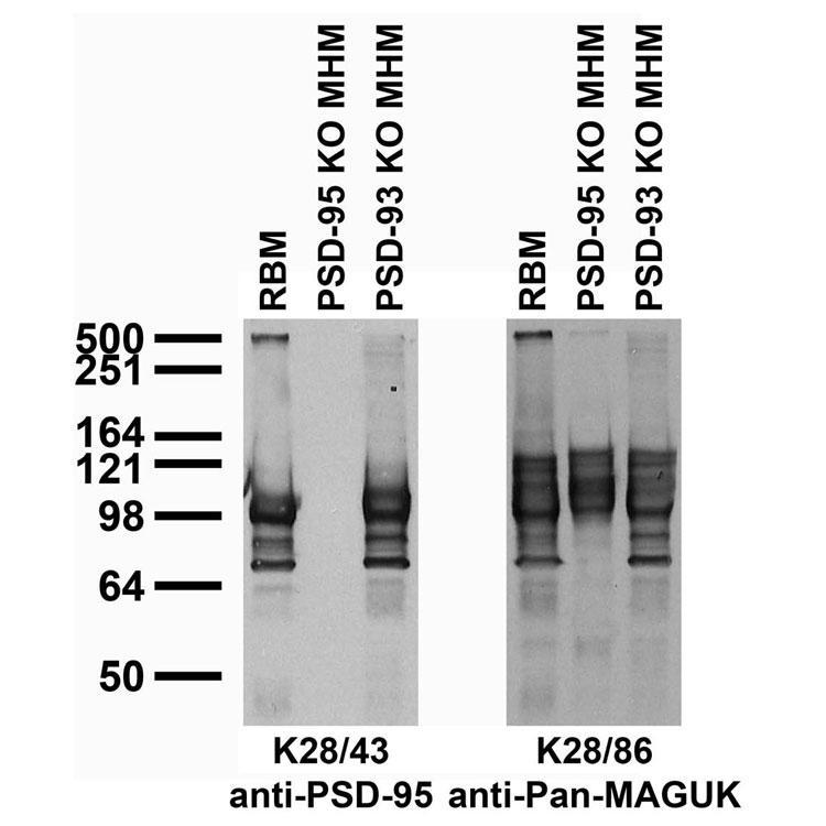
- immunoblot against adult rat brain membranes (RBM) and adult mouse hippocampal membranes (MHM) from PSD-95 and -93 knockout (KO) mice probed with K28/43 (left) or K28/86 (right) TC supe. Mouse samples courtesy of Richard Huganir (Johns Hopkins University, Howard Hughes Medical Institute).
Supportive validation
- Submitted by
- Antibodies Incorporated / NeuroMab (provider)
- Main image
- Experimental details
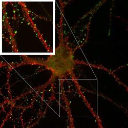
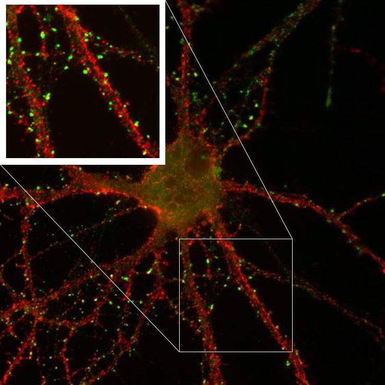
- immunofluorescence staining of cultured rat hippocampal neurons with K28/43 (green) and K57/1 (red, Kv4.2), right image is higher magnification of left image (dotted lines).
Supportive validation
- Submitted by
- Antibodies Incorporated / NeuroMab (provider)
- Main image
- Experimental details
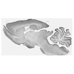
- Adult rat brain immunohistochemistry

Supportive validation
- Submitted by
- Antibodies Incorporated / NeuroMab (provider)
- Main image
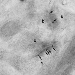
- Experimental details
- electron micrograph of K28/43 hippocampal labelling using a post-embedding immunogold method Immunoparticles (arrows) are seen in the postsynaptic densities of dendritic spines (s) forming asymmetrical synapses with axon terminals (b). Scale bar = 200 nm. Image courtesy of Rafael Lujan (Universidad de Castilla-La Mancha).
