 Explore
 Validate
 Learn
LearnGTX80682
antibody from GeneTex
Targeting: DLG4
PSD-95, PSD95, SAP-90, SAP90
 Western blot Immunocytochemistry
 Immunoprecipitation Immunohistochemistry Flow cytometry Blocking/Neutralizing
Antibody data
- Antibody Data
- Antigen structure
- References [2]
- Comments [0]
- Validations
- Western blot [1]
- Immunocytochemistry [2]
- Flow cytometry [4]
Submit
Validation data
Reference
Comment
Report error
- Product number
- GTX80682 - Provider product page

- Provider
- GeneTex
- Proper citation
- GeneTex Cat#GTX80682, RRID:AB_625474
- Product name
- PSD95 antibody [7E3-1B8]
- Antibody type
- Monoclonal
- Reactivity
- Human, Mouse, Rat, Bovine, Chicken/Avian, Rabbit, Simian, Xenopus, Zebrafish
- Host
- Mouse
Submitted references Diacylglycerol lipaseα (DAGLα) and DAGLβ cooperatively regulate the production of 2-arachidonoyl glycerol in autaptic hippocampal neurons.
Architecture of cannabinoid signaling in mouse retina.
Jain T, Wager-Miller J, Mackie K, Straiker A
Molecular pharmacology 2013 Aug;84(2):296-302
Molecular pharmacology 2013 Aug;84(2):296-302
Architecture of cannabinoid signaling in mouse retina.
Hu SS, Arnold A, Hutchens JM, Radicke J, Cravatt BF, Wager-Miller J, Mackie K, Straiker A
The Journal of comparative neurology 2010 Sep 15;518(18):3848-66
The Journal of comparative neurology 2010 Sep 15;518(18):3848-66
No comments: Submit comment
Supportive validation
- Submitted by
- GeneTex (provider)
- Main image
- Experimental details
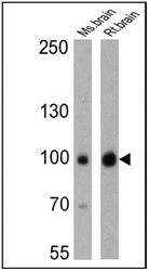
- Western blot analysis of PSD95 was performed by loading 25 ug of mouse brain (lane 1) and rat brain (lane 2) cell lysates onto an SDS polyacrylamide gel. Proteins were transferred to a PVDF membrane and blocked at 4¢XC overnight. The membrane was probed with a PSD95 monoclonal antibody at a dilution of 1:200 overnight at 4¢XC, washed in TBST, and probed with an HRP-conjugated secondary antibody for 1 hr at room temperature in the dark. Chemiluminescent detection was performed using Pierce ECL Plus Western Blotting Substrate. Results show a band at ~95 kDa.

Supportive validation
- Submitted by
- GeneTex (provider)
- Main image
- Experimental details
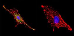
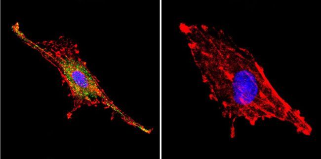
- Immunofluorescent analysis of PSD95 using Anti-PSD95 Monoclonal Antibody (7E3-1B8) shows staining in C6 Cells. PSD95 staining (green), F-Actin staining with Phalloidin (red) and nuclei with DAPI (blue) is shown. Cells were grown on chamber slides and fixed with formaldehyde prior to staining. Cells were probed without (control) or with or an antibody recognizing PSD95 at a dilution of 1:20 over night at 4 ?C, washed with PBS and incubated with a DyLight-488 conjugated secondary antibody. Images were taken at 60X magnification.
- Submitted by
- GeneTex (provider)
- Main image

- Experimental details
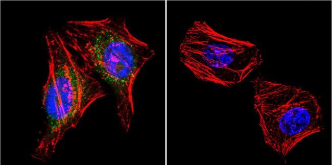
- Immunofluorescent analysis of PSD95 using Anti-PSD95 Monoclonal Antibody (7E3-1B8) shows staining in Hela Cells. PSD95 staining (green), F-Actin staining with Phalloidin (red) and nuclei with DAPI (blue) is shown. Cells were grown on chamber slides and fixed with formaldehyde prior to staining. Cells were probed without (control) or with or an antibody recognizing PSD95 at a dilution of 1:20 over night at 4 ?C, washed with PBS and incubated with a DyLight-488 conjugated secondary antibody. Images were taken at 60X magnification.
Supportive validation
- Submitted by
- GeneTex (provider)
- Main image
- Experimental details
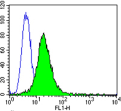
- Flow cytometry analysis of PSD95 showing positive staining in the membrane of Neuro-2a cells compared to an isotype control (blue). Cells were harvested, adjusted to a concentration of 1-5x10^6 cells/ml, fixed with 2% paraformaldehyde and washed with PBS. Cells were blocked with a 2% solution of BSA-PBS for 30 min at room temperature and incubated with a PSD95 monoclonal antibody (GTX80682) at a dilution of 2 ug/test for 60 min at room temperature. Cells were then incubated for 40 min at room temperature in the dark using a Dylight 488-conjugated goat anti-mouse IgG (H+L) secondary antibody and re-suspended in PBS for FACS analysis.
- Validation comment
- FACS

- Submitted by
- GeneTex (provider)
- Main image
- Experimental details
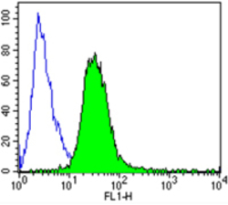
- Flow cytometry analysis of PSD95 showing positive staining in the membrane of SH-SY5Y cells compared to an isotype control (blue). Cells were harvested, adjusted to a concentration of 1-5x10^6 cells/ml, fixed with 2% paraformaldehyde and washed with PBS. Cells were blocked with a 2% solution of BSA-PBS for 30 min at room temperature and incubated with a PSD95 monoclonal antibody (GTX80682) at a dilution of 2 ug/test for 60 min at room temperature. Cells were then incubated for 40 min at room temperature in the dark using a Dylight 488-conjugated goat anti-mouse IgG (H+L) secondary antibody and re-suspended in PBS for FACS analysis.
- Validation comment
- FACS

- Submitted by
- GeneTex (provider)
- Main image
- Experimental details
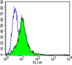
- Flow cytometry analysis of PSD95 showing positive staining in the membrane of U87-MG cells compared to an isotype control (blue). Cells were harvested, adjusted to a concentration of 1-5x10^6 cells/ml, fixed with 2% paraformaldehyde and washed with PBS. Cells were blocked with a 2% solution of BSA-PBS for 30 min at room temperature and incubated with a PSD95 monoclonal antibody (GTX80682) at a dilution of 2 ug/test for 60 min at room temperature. Cells were then incubated for 40 min at room temperature in the dark using a Dylight 488-conjugated goat anti-mouse IgG (H+L) secondary antibody and re-suspended in PBS for FACS analysis.
- Validation comment
- FACS
- Submitted by
- GeneTex (provider)
- Main image
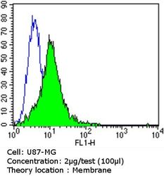
- Experimental details
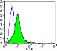
- Flow cytometry analysis of PSD95 in U87-MG cells compared to an isotype control (blue). Cells were harvested, adjusted to a concentration of 1-5x10^6 cells/ml, fixed with 2% paraformaldehyde and washed with PBS. Cells were blocked with a 2% solution of BSA-PBS for 30 min at room temperature and incubated with a PSD95 monoclonal antibody at a dilution of 2 ug/test for 60 min at room temperature. Cells were then incubated for 40 min at room temperature in the dark using a Dylight 488-conjugated goat anti-mouse IgG (H+L) secondary antibody and re-suspended in PBS for FACS analysis.
