Explore
 Validate
 Learn
Learn Western blot
Antibody data
- Antibody Data
- Antigen structure
- References [0]
- Comments [0]
- Validations
- Western blot [3]
- Immunocytochemistry [1]
- Immunohistochemistry [1]
Submit
Validation data
Reference
Comment
Report error
- Product number
- 710179 - Provider product page

- Provider
- Invitrogen Antibodies
- Product name
- Adiponectin Recombinant Polyclonal Antibody
- Antibody type
- Polyclonal
- Antigen
- Recombinant full-length protein
- Reactivity
- Human, Mouse, Rat
- Host
- Rabbit
- Isotype
- IgG
- Vial size
- 100 µg
- Concentration
- 0.5 mg/mL
- Storage
- Store at 4°C short term. For long term storage, store at -20°C, avoiding freeze/thaw cycles.
No comments: Submit comment
Supportive validation
- Submitted by
- Invitrogen Antibodies (provider)
- Main image
- Experimental details
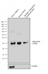
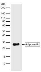
- Western blot analysis was performed on tissue extract (30 µg lysate) of Mouse Adipose (Lane 1), 2 µL of Human Plasma (Lane 2) and 2 µL of conditioned media from BM-MSC differentiated into adipocytes (Lane 3). The blot was probed with Anti-Adiponectin Recombinant Rabbit Polyclonal Antibody (Product # 710179, 2 µg/mL) and detected by chemiluminescence using Goat anti-Rabbit IgG (H+L) Superclonal™ Secondary Antibody, HRP conjugate (Product # A27036, 0.25 µg/mL, 1:4000 dilution). A 26 kDa band corresponding to Adiponectin was observed across the samples tested. Known quantity of protein samples were electrophoresed using Novex®NuPAGE®4-12 % Bis-Tris gel (Product # NP0321BOX), XCell SureLock™ Electrophoresis System (Product # EI0002) and Novex® Sharp Pre-Stained Protein Standard (Product # LC5800). Resolved proteins were then transferred onto a nitrocellulose membrane using the wet transfer system. The membrane was probed with the relevant primary and secondary antibody following blocking with 5 % skimmed milk. Chemiluminescent detection was performed using Pierce™ ECL Western blotting Substrate (Product # 32106).

- Submitted by
- Invitrogen Antibodies (provider)
- Main image
- Experimental details
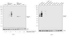
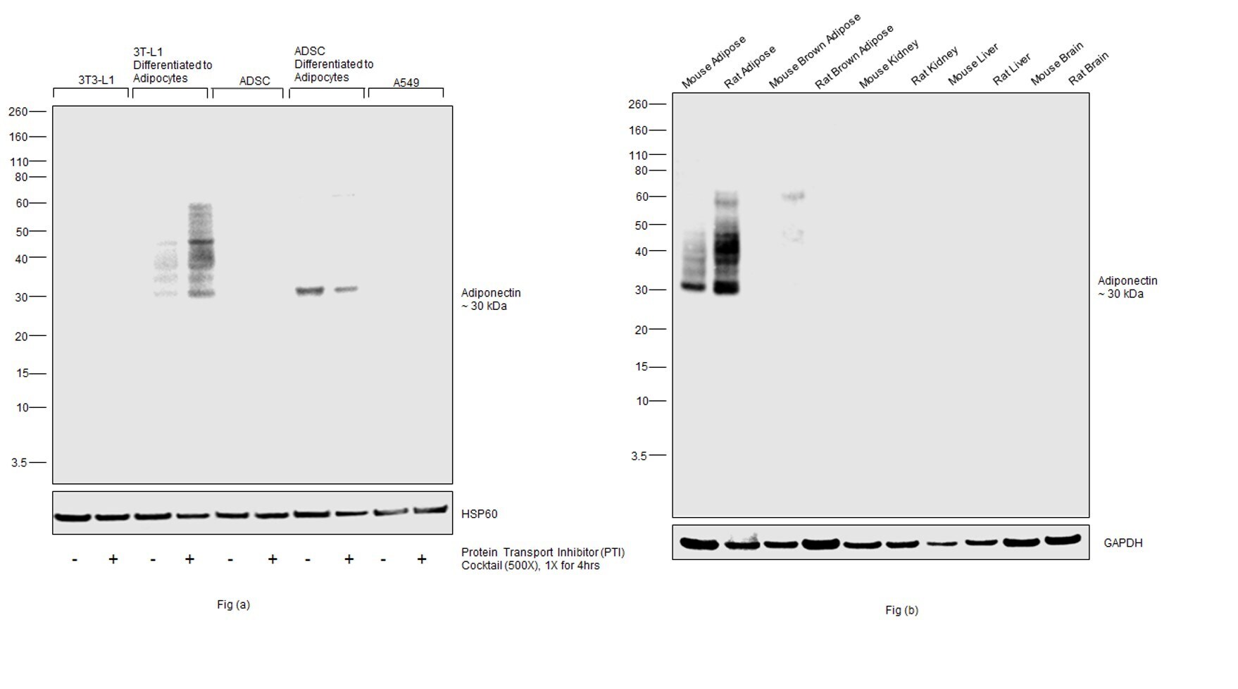
- Western blot was performed using Anti-Adiponectin (Product # 710179) and a 30 kDa band corresponding to Adiponectin was observed across cell lines and tissues tested. Whole cell extracts (30 µg lysate) of (Fig. a) 3T3-L1 (Lane 1), 3T3-L1 treated with PTI (1X, 4hrs) (Lane 2), 3T3-L1 differentiated to Adipocytes (Lane 3), 3T3-L1 differentiated to Adipocytes further treated with PTI (1X, 4hrs) (Lane 4), ADSC (Lane 5), ADSC treated with PTI (1X, 4hrs) (Lane 6), ADSC differentiated to Adipocytes (Lane 7), ADSC differentiated to Adipocytes further treated with PTI (1X, 4hrs) (Lane 8) and A549 (Lane 9), A549 treated with PTI (1X, 4hrs) (Lane 10); (Fig. b) Mouse Adipose (Lane 1), Rat Adipose (Lane 2), Mouse Brown Adipose (Lane 3), Rat Brown Adipose (Lane 4), Mouse Kidney (Lane 5), Rat Kidney (Lane 6), Mouse Liver (Lane 7), Rat Liver (Lane 8), Mouse Brain (Lane 9) and Rat Brain (Lane 10) were electrophoresed using NuPAGE™ 4-12% Bis-Tris Protein Gel (Product # NP0321BOX). Resolved proteins were then transferred onto a nitrocellulose membrane (Product # IB23002) by iBlot® 2 Dry Blotting System (Product # IB21001). The blot was probed with the primary antibody (1 µg/mL) and detected by chemiluminescence with Goat anti-Rabbit IgG (H+L) Superclonal™ Recombinant Secondary Antibody, HRP (Product # A27036,1:20000 dilution) using the iBright FL 1000 (Product # A32752). Chemiluminescent detection was performed using SuperSignal™ West Pico PLUS Chemiluminescent Subst
- Submitted by
- Invitrogen Antibodies (provider)
- Main image
- Experimental details
- Western blot analysis of Adiponectin in whole cell extracts from serum-starved 3T3 L1 cells treated with Insulin (100 ng/mL, 15 min) using an Adiponectin Recombinant Rabbit Polyclonal Antibody (Product # 710179) at a dilution of 1 µg/mL. Samples were detected using chemiluminescence (ECL). Results show a band at ~28 kDa.

Supportive validation
- Submitted by
- Invitrogen Antibodies (provider)
- Main image

- Experimental details
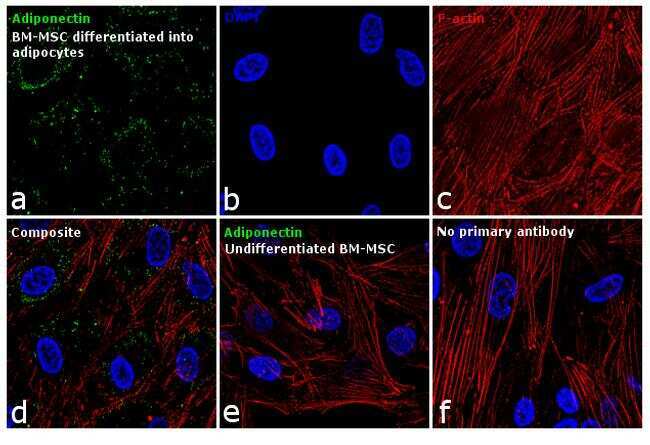
- Immunofluorescence analysis of Adiponectin was performed using 90% confluent BM-MSCs differentiated into adipocytes. The cells were fixed with 4% paraformaldehyde for 10 minutes, permeabilized with 0.1% Triton™ X-100 for 10 minutes, and blocked with 1% BSA for 1 hour at room temperature. The cells were labeled with Adiponectin Antibody (11HCLC) Recombinant Rabbit Polyclonal (Product # 710179) at 5 µg/mL in 0.1% BSA and incubated overnight at 4 degree Celsius and then labeled with Goat anti-Rabbit IgG (H+L) Superclonal™ Secondary Antibody, Alexa Fluor® 488 conjugate (Product # A27034) at a dilution of 1:2000 for 45 minutes at room temperature (Panel a: green). Nuclei (Panel b: blue) were stained with SlowFade® Gold Antifade Mountant with DAPI (Product # S36938). F-actin (Panel c: red) was stained with Rhodamine Phalloidin (Product # R415, 1:300). Panel d represents the merged image showing cytoplasmic as well as extracellular localization. Panel e represents undifferentiated BM-MSCs showing no Adiponectin staining. Panel f represents control cells with no primary antibody to assess background. The images were captured at 60X magnification.
Supportive validation
- Submitted by
- Invitrogen Antibodies (provider)
- Main image
- Experimental details
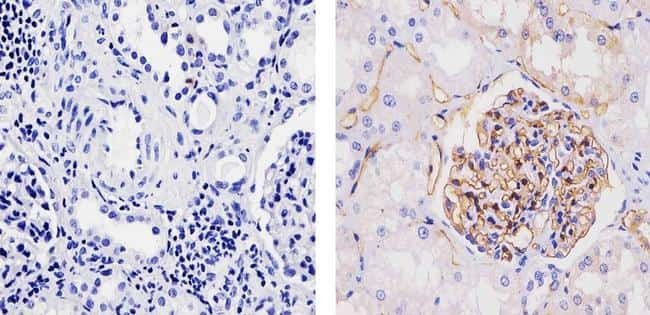
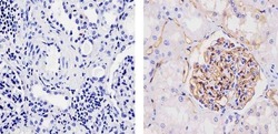
- Immunohistochemistry analysis of Adiponectin showing staining in the membrane of paraffin-embedded human kidney tissue (right) compared to a negative control without primary antibody (left). To expose target proteins, antigen retrieval was performed using 10mM sodium citrate (pH 6.0), microwaved for 8-15 min. Following antigen retrieval, tissues were blocked in 3% H2O2-methanol for 15 min at room temperature, washed with ddH2O and PBS, and then probed with a Adiponectin Recombinant Rabbit Polyclonal Antibody (Product # 710179) diluted in 3% BSA-PBS at a dilution of 1:100 for 1 hour at 37ºC in a humidified chamber. Tissues were washed extensively in PBST and detection was performed using an HRP-conjugated secondary antibody followed by colorimetric detection using a DAB kit. Tissues were counterstained with hematoxylin and dehydrated with ethanol and xylene to prep for mounting.