Explore
 Validate
 Learn
Learn14-4040-82
antibody from Invitrogen Antibodies
Targeting: TRAF3IP2
ACT1, C6orf2, C6orf4, C6orf5, C6orf6, CIKS, DKFZP586G0522
 Western blot
 Other assay
Antibody data
- Antibody Data
- Antigen structure
- References [7]
- Comments [0]
- Validations
- Other assay [3]
Submit
Validation data
Reference
Comment
Report error
- Product number
- 14-4040-82 - Provider product page

- Provider
- Invitrogen Antibodies
- Product name
- ACT1 Monoclonal Antibody (9ACT12), eBioscience™
- Antibody type
- Monoclonal
- Antigen
- Other
- Description
- Description: The monoclonal antibody 9ACT12 reacts with human Act1 (NF-kappaB activator 1), also known as CIKS (a connection to IkB kinase and stress-activated kinase). Originally cloned for its ability to activate NF-kappaB and its direct interaction with IKKgamma, Act1 has been subsequently shown to play several roles in immunity. In B cells, it is a negative regulator of CD40 and BAFFR signaling affecting reduced survival. This regulation is through direct interaction of Act1 with CD40 or BAFFR and TRAF3. In addition, Act1 is involved in IL-17R signaling. It interacts with IL-17RA through the homotypic SEFIR interaction domain and is critical for downstream NF-kappaB- and C/EBP-mediated inflammatory responses. Applications Reported: This 9ACT12 antibody has been reported for use in immunoblotting (WB). Applications Tested: This 9ACT12 antibody has been tested by immunoblot of human peripheral blood cell lysates. This can be used at less than or equal to 1-5 µg/mL. It is recommended that the antibody be carefully titrated for optimal performance in the assay of interest. Purity: Greater than 90%, as determined by SDS-PAGE. Aggregation: Less than 10%, as determined by HPLC. Filtration: 0.2 µm post-manufacturing filtered.
- Reactivity
- Human
- Host
- Rat
- Isotype
- IgG
- Antibody clone number
- 9ACT12
- Vial size
- 100 μg
- Concentration
- 0.5 mg/mL
- Storage
- 4°C
Submitted references Fusion transcripts FYN-TRAF3IP2 and KHDRBS1-LCK hijack T cell receptor signaling in peripheral T-cell lymphoma, not otherwise specified.
Natural killer cell functional defects in pediatric patients with severe and recurrent herpesvirus infections.
IL-17 receptor adaptor protein Act1/CIKS plays an evolutionarily conserved role in antiviral signaling.
IL-17 receptor signaling inhibits C/EBPbeta by sequential phosphorylation of the regulatory 2 domain.
The critical role of epithelial-derived Act1 in IL-17- and IL-25-mediated pulmonary inflammation.
The adaptor protein CIKS/Act1 is essential for IL-25-mediated allergic airway inflammation.
Deficiency of Act1, a critical modulator of B cell function, leads to development of Sjögren's syndrome.
Debackere K, Marcelis L, Demeyer S, Vanden Bempt M, Mentens N, Gielen O, Jacobs K, Broux M, Verhoef G, Michaux L, Graux C, Wlodarska I, Gaulard P, de Leval L, Tousseyn T, Cools J, Dierickx D
Nature communications 2021 Jun 17;12(1):3705
Nature communications 2021 Jun 17;12(1):3705
Natural killer cell functional defects in pediatric patients with severe and recurrent herpesvirus infections.
Ornstein BW, Hill EB, Geurs TL, French AR
The Journal of infectious diseases 2013 Feb 1;207(3):458-68
The Journal of infectious diseases 2013 Feb 1;207(3):458-68
IL-17 receptor adaptor protein Act1/CIKS plays an evolutionarily conserved role in antiviral signaling.
Ryzhakov G, Blazek K, Lai CC, Udalova IA
Journal of immunology (Baltimore, Md. : 1950) 2012 Nov 15;189(10):4852-8
Journal of immunology (Baltimore, Md. : 1950) 2012 Nov 15;189(10):4852-8
IL-17 receptor signaling inhibits C/EBPbeta by sequential phosphorylation of the regulatory 2 domain.
Shen F, Li N, Gade P, Kalvakolanu DV, Weibley T, Doble B, Woodgett JR, Wood TD, Gaffen SL
Science signaling 2009 Feb 24;2(59):ra8
Science signaling 2009 Feb 24;2(59):ra8
The critical role of epithelial-derived Act1 in IL-17- and IL-25-mediated pulmonary inflammation.
Swaidani S, Bulek K, Kang Z, Liu C, Lu Y, Yin W, Aronica M, Li X
Journal of immunology (Baltimore, Md. : 1950) 2009 Feb 1;182(3):1631-40
Journal of immunology (Baltimore, Md. : 1950) 2009 Feb 1;182(3):1631-40
The adaptor protein CIKS/Act1 is essential for IL-25-mediated allergic airway inflammation.
Claudio E, Sønder SU, Saret S, Carvalho G, Ramalingam TR, Wynn TA, Chariot A, Garcia-Perganeda A, Leonardi A, Paun A, Chen A, Ren NY, Wang H, Siebenlist U
Journal of immunology (Baltimore, Md. : 1950) 2009 Feb 1;182(3):1617-30
Journal of immunology (Baltimore, Md. : 1950) 2009 Feb 1;182(3):1617-30
Deficiency of Act1, a critical modulator of B cell function, leads to development of Sjögren's syndrome.
Qian Y, Giltiay N, Xiao J, Wang Y, Tian J, Han S, Scott M, Carter R, Jorgensen TN, Li X
European journal of immunology 2008 Aug;38(8):2219-28
European journal of immunology 2008 Aug;38(8):2219-28
No comments: Submit comment
Supportive validation
- Submitted by
- Invitrogen Antibodies (provider)
- Main image

- Experimental details
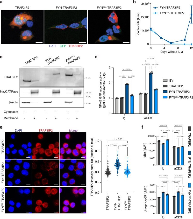
- Fig. 3 Membrane anchoring of FYN-TRAF3IP2 is required to activate NF-kappaB signaling. a Immunofluorescence images of 293T cells transfected with TRAF3IP2 , FYN-TRAF3IP2 and FYN G2A -TRAF3IP2 stained for TRAF3IP2. Scalebars represent 10 um. b Outgrowth of Ba/F3 cells transduced with pMIG- FYN-TRAF3IP2 or pMIG- FYN G2A -TRAF3IP2 after withdrawal of IL-3. n = 3 biological replicates per condition. c Western blot analysis for TRAF3IP2 on cytoplasmic and membrane fractions of Jurkat cells transduced with pMIG- TRAF3IP2 , pMIG- FYN-TRAF3IP2 and pMIG- FYN G2A -TRAF3IP2 . d NF-kappaB GFP reporter signal intensity in Jurkat cells with empty control vector or expression of TRAF3IP2, FYN-TRAF3IP2 or FYN G2A -TRAF3IP2 after stimulation with an agonistic anti-CD3epsilon antibody or exposure to control immunoglobulin (Ig). n = 3 biological replicates per condition for Ig control, n = 4 for anti-CD3epsilon. p values were calculated with Tukey's post-hoc multiple comparisons test. e Immunofluorescence images (left) of TRAF3IP2 staining on CD4 + T cells transduced to express TRAF3IP2, FYN-TRAF3IP2 or FYN G2A -TRAF3IP2. Scalebars represent 5 um. Quantification (right) of TRAF3IP2 signal intensity in the outer cell bin, expressed as fraction of signal intensity over the entire cell. Each dot represents a cell ( n = 201 for TRAF3IP2, n = 192 for FYN-TRAF3IP2 and n = 179 for FYN G2A -TRAF3IP2). Horizontal line and whiskers represent median and interquartile range respectively. p values were calc
- Submitted by
- Invitrogen Antibodies (provider)
- Main image
- Experimental details
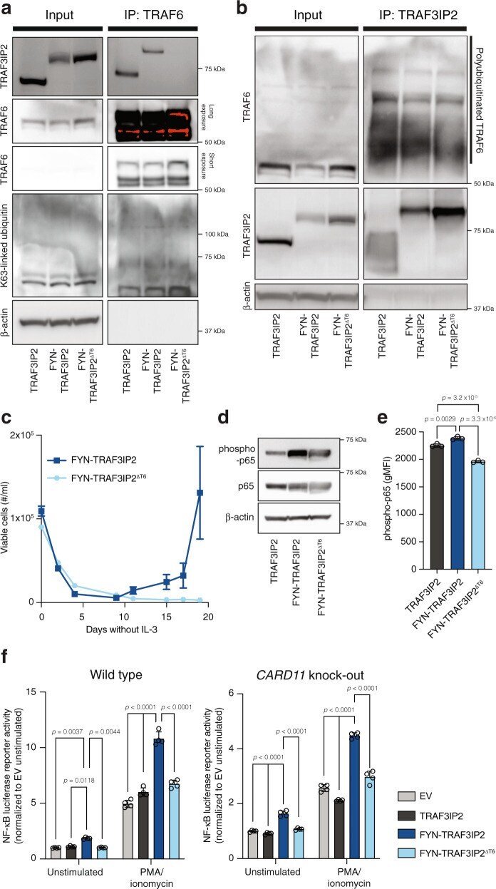
- Fig. 4 FYN-TRAF3IP2 activates TRAF6 and canonical NF-kappaB signaling independent of the CBM signalosome. a Western blot analysis of whole-cell lysate and anti-TRAF6 immunoprecipitate in TRAF3IP2-, FYN-TRAF3IP2- and FYN-TRAF3IP2 DeltaT6 -expressing Jurkat cells. b Western blot analysis of whole-cell lysate and anti-TRAF3IP2 immunoprecipitate in TRAF3IP2-, FYN-TRAF3IP2- and FYN-TRAF3IP2 DeltaT6 -expressing Jurkat cells. c Outgrowth of Ba/F3 cells transduced with pMIG- FYN-TRAF3IP2 or pMIG- FYN-TRAF3IP2 DeltaT6 after withdrawal of IL-3. n = 3 biological replicates per condition. d Western blot analysis for canonical NF-kappaB activation in Jurkat cells transduced with pMIG- TRAF3IP2 , pMIG- FYN-TRAF3IP2 or pMIG- FYN-TRAF3IP2 DeltaT6 . e Intracellular flow cytometry for phospho-p65 in resting CD4 + T cells with expression of TRAF3IP2, FYN-TRAF3IP2 or FYN-TRAF3IP2 DeltaT6 . n = 3 biological replicates per condition. p values were calculated with Tukey's post-hoc multiple comparisons test. f NF-kappaB luciferase reporter signal intensity in wild-type Jurkat cells (left) or CARD11 knock-out Jurkat cells (right) with empty control vector or expression of TRAF3IP2, FYN-TRAF3IP2 or FYN-TRAF3IP2 DeltaT6 in resting conditions or after stimulation with PMA. n = 4 biological replicates per condition. p values were calculated with Tukey's post-hoc multiple comparisons test. All data are represented as mean +- SD.
- Submitted by
- Invitrogen Antibodies (provider)
- Main image

- Experimental details
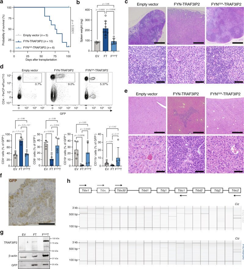
- Fig. 5 Expression of FYN-TRAF3IP2 in hematopoietic cells fosters the development of PTCL-NOS. a Kaplan-Meier survival curve after transplantation with HSPC transduced with empty pMIG vector ( n = 5), pMIG- FYN-TRAF3IP2 ( n = 10) or pMIG- FYN G2A -TRAF3IP2-GFP ( n = 6). Log-rank p value was obtained from a two-sided Chi-square test. b Spleen weights at sacrifice of mice transplanted with HSPC transduced with empty vector (EV, n = 5), FYN-TRAF3IP2 (FT, n = 9) or FYN G2A -TRAF3IP2 (F G2A T, n = 6). p values were calculated with Tukey's post-hoc multiple comparisons test. c Representative H&E stains of lymph nodes from mice transplanted with HSPC transduced with empty vector ( n = 5), FYN-TRAF3IP2 ( n = 5) or FYN G2A -TRAF3IP2 ( n = 5). Low magnification images are in the top row, scalebars represent 500 um. High magnification images are in the bottom row, scalebars represent 50 um. d Representative flow cytometry plots for cell suspensions from the lymph nodes of EV mice, FT mice and F G2A T mice (top). Quantification of the lineage of GFP + cells in lymph node cell suspensions (bottom). n = 5 mice per group. p values were calculated with Tukey's post-hoc multiple comparisons test. e Representative H&E stains of livers from mice transplanted with HSPC transduced with empty vector ( n = 5), FYN-TRAF3IP2 ( n = 5) or FYN G2A -TRAF3IP2 ( n = 5). Low magnification images are in the top row, scalebars represent 500 um. High magnification images are in the bottom row, scalebars represe