Explore
 Validate
 Learn
Learn Western blot
 Immunohistochemistry
Antibody data
- Antibody Data
- Antigen structure
- References [0]
- Comments [0]
- Validations
- Immunohistochemistry [2]
Submit
Validation data
Reference
Comment
Report error
- Product number
- PA3-038 - Provider product page

- Provider
- Invitrogen Antibodies
- Product name
- GPR61 Polyclonal Antibody
- Antibody type
- Polyclonal
- Antigen
- Other
- Description
- IHC(P) analysis shows positive staining of GPR61 in human pancreatic islets and enteric ganglion. WB analysis shows GPR61 in glyco-protein enriched fractions from mock transfected or GPR61 overexpressed 293 cells. The GPR61 antiserum showed specific detection of GPR61 over other tested GPRs. GPR61 exists in different glycosylated forms and can form dimers and oligomers. Consequently it appears as a long smear on WB or is detected at multiple molecular weights.
- Reactivity
- Human
- Host
- Rabbit
- Isotype
- IgG
- Vial size
- 100 μL
- Concentration
- Conc. Not Determined
- Storage
- -20°C, Avoid Freeze/Thaw Cycles
No comments: Submit comment
Supportive validation
- Submitted by
- Invitrogen Antibodies (provider)
- Main image
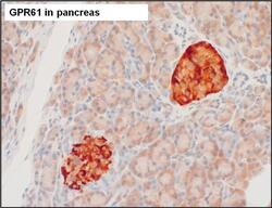
- Experimental details
- Immunohistochemistry analysis of GPR61 was performed on human pancreas tissue. To expose target proteins, antigen retrieval was performed by microwaving tissues for 20 minutes in 10mM sodium citrate buffer (pH 6.0). Tissue slides were probed with a GPR61 polyclonal antibody (Product # PA3-038) at a dilution of 1:3000, overnight at 4C in a humidified chamber. Tissues were washed, and detection was performed using an ABC kit composed of biotinylated goat anti-rabbit IgG, peroxidase-conjugated avidin, and 3-amino-9-ethylcarbazole (AEC) substrate in acetate buffer. Tissues were counterstained with hematoxylin and dehydrated to prep for mounting.

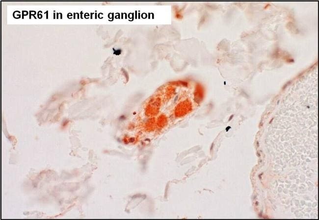
- Submitted by
- Invitrogen Antibodies (provider)
- Main image
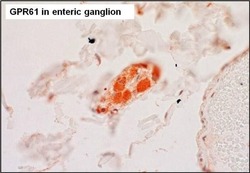
- Experimental details
- Immunohistochemistry analysis of GPR61 was performed on human enteric ganglion tissue. To expose target proteins, antigen retrieval was performed by microwaving tissues for 20 minutes in 10mM sodium citrate buffer (pH 6.0). Tissue slides were probed with a GPR61 polyclonal antibody (Product # PA3-038) at a dilution of 1:3000, overnight at 4C in a humidified chamber. Tissues were washed, and detection was performed using an ABC kit composed of biotinylated goat anti-rabbit IgG, peroxidase-conjugated avidin, and 3-amino-9-ethylcarbazole (AEC) substrate in acetate buffer. Tissues were counterstained with hematoxylin and dehydrated to prep for mounting.