Explore
 Validate
 Learn
Learn Western blot
 ELISA
ELISAAntibody data
- Antibody Data
- Antigen structure
- References [1]
- Comments [0]
- Validations
- Western blot [3]
- Immunocytochemistry [1]
Submit
Validation data
Reference
Comment
Report error
- Product number
- MA1-20581 - Provider product page

- Provider
- Invitrogen Antibodies
- Product name
- RAN Monoclonal Antibody (ARAN1)
- Antibody type
- Monoclonal
- Antigen
- Recombinant full-length protein
- Description
- Recommended positive controls: Jurkat.
- Reactivity
- Human, Mouse, Rat, Bovine, Hamster, Xenopus
- Host
- Mouse
- Isotype
- IgG
- Antibody clone number
- ARAN1
- Vial size
- 50 µL
- Concentration
- 2.3 mg/mL
- Storage
- Store at 4°C short term. For long term storage, store at -20°C, avoiding freeze/thaw cycles.
Submitted references Neuroprotection resulting from insufficiency of RANBP2 is associated with the modulation of protein and lipid homeostasis of functionally diverse but linked pathways in response to oxidative stress.
Cho KI, Yi H, Tserentsoodol N, Searle K, Ferreira PA
Disease models & mechanisms 2010 Sep-Oct;3(9-10):595-604
Disease models & mechanisms 2010 Sep-Oct;3(9-10):595-604
No comments: Submit comment
Supportive validation
- Submitted by
- Invitrogen Antibodies (provider)
- Main image

- Experimental details
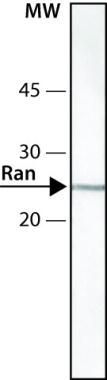
- Western blot of RAN in whole extract of Jurkat cells using a RAN monoclonal antibody (Product # MA1-20581) at a dilution of 0.5 µg/mL and developed using a Goat anti-mouse IgG-ALP and NBT/BCIP substrate.
- Submitted by
- Invitrogen Antibodies (provider)
- Main image
- Experimental details
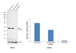
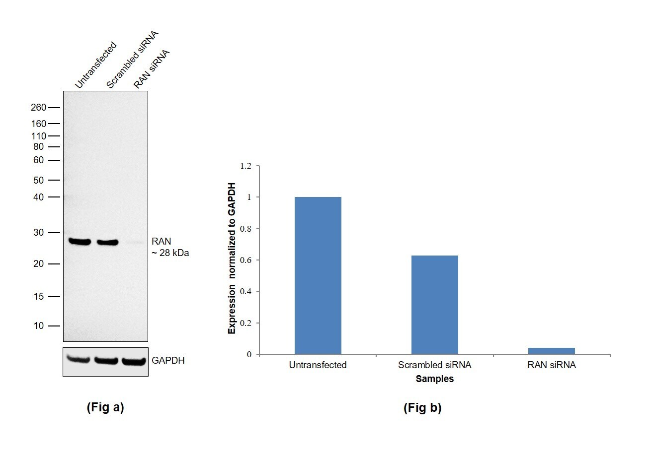
- Knockdown of RAN was achieved by transfecting HeLa with RAN specific siRNA (Silencer® select Product # s11769). Western blot analysis (Fig. a) was performed using whole cell extracts from the RAN knockdown cells (lane 3), non-specific scrambled siRNA transfected cells (lane 2) and untransfected cells (lane 1). The blot was probed with RAN Monoclonal Antibody (Product # MA1-20581, 0.5 µg/ml) and Goat anti-Mouse IgG (H+L) Superclonal™ Recombinant Secondary Antibody, HRP (Product # A28177, 0.25µg/ml, 1:4000 dilution). Densitometric analysis of this western blot is shown in histogram (Fig. b). Decrease in signal upon siRNA mediated knock down confirms that antibody is specific to RAN.
- Submitted by
- Invitrogen Antibodies (provider)
- Main image
- Experimental details
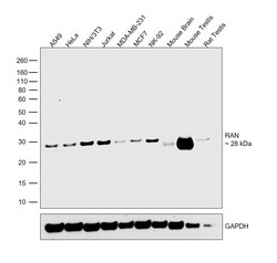
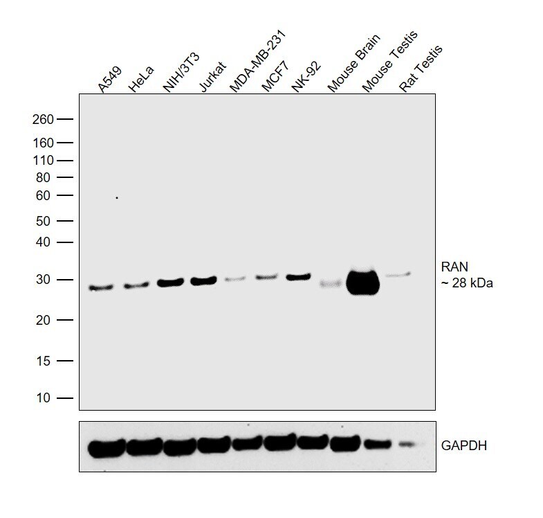
- Western blot was performed using Anti-RAN Monoclonal Antibody (Product # MA1-20581) and a 28 kDa band corresponding to RAN was observed across all the cell lines and tissues tested. Whole cell extracts (30 µg lysate) of A549 (Lane 1), HeLa (Lane 2), NIH/3T3 (Lane 3), Jurkat (Lane 4), MDA-MB-231 (Lane 5), MCF7 (Lane 6), NK-92 (Lane 7), tissue extracts of Mouse Brain (Lane 8), Mouse Testis (Lane 9) and Rat Testis (Lane 10) were electrophoresed using NuPAGE™ 10% Bis-Tris Protein Gel (Product # NP0302BOX). Resolved proteins were then transferred onto a nitrocellulose membrane (Product # IB23001) by iBlot® 2 Dry Blotting System (Product # IB21001). The blot was probed with the primary antibody (0.5 µg/ml) and detected by chemiluminescence with Goat anti-Mouse IgG (H+L) Superclonal™ Recombinant Secondary Antibody, HRP (Product # A28177, 1:4000 dilution) using the iBright FL 1000 (Product # A32752). Chemiluminescent detection was performed using Novex® ECL Chemiluminescent Substrate Reagent Kit (Product # WP20005).
Supportive validation
- Submitted by
- Invitrogen Antibodies (provider)
- Main image
- Experimental details
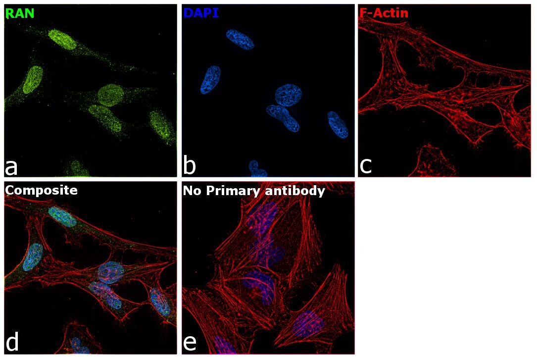
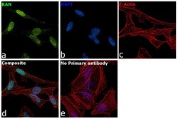
- Immunofluorescence analysis of RAN was performed using 70% confluent log phase HeLa cells. The cells were fixed with 4% paraformaldehyde for 10 minutes, permeabilized with 0.1% Triton™ X-100 for 15 minutes, and blocked with 2% BSA for 1 hour at room temperature. The cells were labeled with RAN Monoclonal Antibody (ARAN1) (Product # MA1-20581) at 5 µg/mL in 0.1% BSA, incubated at 4 degree celsius overnight and then with Donkey anti-Mouse IgG (H+L) Highly Cross-Adsorbed Secondary Antibody, Alexa Fluor Plus 488 (Product # A32766) at a dilution of 1:2000 for 45 minutes at room temperature (Panel a: green). Nuclei (Panel b: blue) were stained with SlowFade® Gold Antifade Mountant with DAPI (Product # S36938). F-actin (Panel c: red) was stained with Rhodamine Phalloidin (Product # R415, 1:300). Panel d represents the merged image showing staining in nucleus and cytoplasm. Panel e represents control cells with no primary antibody to assess background. The images were captured at 60X magnification.