Explore
 Validate
 Learn
Learn Western blot
Antibody data
- Antibody Data
- Antigen structure
- References [2]
- Comments [0]
- Validations
- Western blot [1]
- Immunohistochemistry [1]
Submit
Validation data
Reference
Comment
Report error
- Product number
- AF3317 - Provider product page

- Provider
- R&D Systems
- Product name
- Human HES-1 Antibody
- Antibody type
- Polyclonal
- Description
- Immunogen affinity purified. Detects human HES-1 in direct ELISAs and Western blots. In direct ELISAs, less than 2% cross-reactivity with recombinant human (rh) HES-2, rhHES-4, rhHES-5, rhHES-6, and rhHES-7 is observed.
- Reactivity
- Human
- Host
- Goat
- Conjugate
- Unconjugated
- Antigen sequence
Q14469- Isotype
- IgG
- Vial size
- 100 ug
- Concentration
- LYOPH
- Storage
- Use a manual defrost freezer and avoid repeated freeze-thaw cycles. 12 months from date of receipt, -20 to -70 °C as supplied. 1 month, 2 to 8 °C under sterile conditions after reconstitution. 6 months, -20 to -70 °C under sterile conditions after reconstitution.
Submitted references Interplay between CCR7 and Notch1 axes promotes stemness in MMTV-PyMT mammary cancer cells.
Rabconnectin-3 is a functional regulator of mammalian Notch signaling.
Boyle ST, Gieniec KA, Gregor CE, Faulkner JW, McColl SR, Kochetkova M
Molecular cancer 2017 Jan 31;16(1):19
Molecular cancer 2017 Jan 31;16(1):19
Rabconnectin-3 is a functional regulator of mammalian Notch signaling.
Sethi N, Yan Y, Quek D, Schupbach T, Kang Y
The Journal of biological chemistry 2010 Nov 5;285(45):34757-64
The Journal of biological chemistry 2010 Nov 5;285(45):34757-64
No comments: Submit comment
Supportive validation
- Submitted by
- R&D Systems (provider)
- Main image
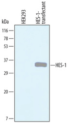
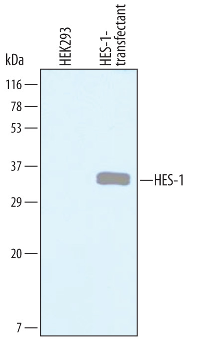
- Experimental details
- Detection of Human HES-1 by Western Blot. Western blot shows lysates of HEK293 human embryonic kidney cell line either mock transfected or transfected with human HES-1. PVDF Membrane was probed with 1 µg/mL of Goat Anti-Human HES-1 Antigen Affinity-purified Polyclonal Antibody (Catalog # AF3317) followed by HRP-conjugated Anti-Goat IgG Secondary Antibody (Catalog # HAF019). A specific band was detected for HES-1 at approximately 35 kDa (as indicated). This experiment was conducted under reducing conditions and using Immunoblot Buffer Group 8.
Supportive validation
- Submitted by
- R&D Systems (provider)
- Main image

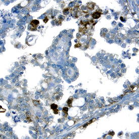
- Experimental details
- HES-1 in Human Ovarian Cancer Tissue. HES-1 was detected in immersion fixed paraffin-embedded sections of human ovarian cancer tissue using Goat Anti-Human HES-1 Antigen Affinity-purified Polyclonal Antibody (Catalog # AF3317) at 15 µg/mL overnight at 4 °C. Tissue was stained using the Anti-Goat HRP-DAB Cell & Tissue Staining Kit (brown; Catalog # CTS008) and counterstained with hematoxylin (blue). Specific labeling was localized to the cytoplasm of cancer cells. View our protocol for Chromogenic IHC Staining of Paraffin-embedded Tissue Sections.