Explore
 Validate
 Learn
Learn Western blot
 Immunocytochemistry
Antibody data
- Antibody Data
- Antigen structure
- References [3]
- Comments [0]
- Validations
- Immunocytochemistry [4]
- Other assay [5]
Submit
Validation data
Reference
Comment
Report error
- Product number
- PA5-42883 - Provider product page

- Provider
- Invitrogen Antibodies
- Product name
- ACSL3 Polyclonal Antibody
- Antibody type
- Polyclonal
- Antigen
- Synthetic peptide
- Description
- Peptide sequence: LDGLASVLYP GCDTLDKVFT YAKNKFKNKR LLGTREVLNE EDEVQPNGKI Sequence homology: Cow: 93%; Dog: 86%; Guinea Pig: 93%; Horse: 93%; Human: 100%; Mouse: 100%; Rabbit: 93%; Rat: 93%
- Reactivity
- Human
- Host
- Rabbit
- Isotype
- IgG
- Vial size
- 100 μL
- Concentration
- 0.5 mg/mL
- Storage
- -20°C, Avoid Freeze/Thaw Cycles
Submitted references TNF-α induces acyl-CoA synthetase 3 to promote lipid droplet formation in human endothelial cells.
Metabolic enzyme ACSL3 is a prognostic biomarker and correlates with anticancer effectiveness of statins in non-small cell lung cancer.
Immunohistochemical staining reveals differential expression of ACSL3 and ACSL4 in hepatocellular carcinoma and hepatic gastrointestinal metastases.
Jung HS, Shimizu-Albergine M, Shen X, Kramer F, Shao D, Vivekanandan-Giri A, Pennathur S, Tian R, Kanter JE, Bornfeldt KE
Journal of lipid research 2020 Jan;61(1):33-44
Journal of lipid research 2020 Jan;61(1):33-44
Metabolic enzyme ACSL3 is a prognostic biomarker and correlates with anticancer effectiveness of statins in non-small cell lung cancer.
Fernández LP, Merino M, Colmenarejo G, Moreno-Rubio J, Sánchez-Martínez R, Quijada-Freire A, Gómez de Cedrón M, Reglero G, Casado E, Sereno M, Ramírez de Molina A
Molecular oncology 2020 Dec;14(12):3135-3152
Molecular oncology 2020 Dec;14(12):3135-3152
Immunohistochemical staining reveals differential expression of ACSL3 and ACSL4 in hepatocellular carcinoma and hepatic gastrointestinal metastases.
Ndiaye H, Liu JY, Hall A, Minogue S, Morgan MY, Waugh MG
Bioscience reports 2020 Apr 30;40(4)
Bioscience reports 2020 Apr 30;40(4)
No comments: Submit comment
Supportive validation
- Submitted by
- Invitrogen Antibodies (provider)
- Main image

- Experimental details
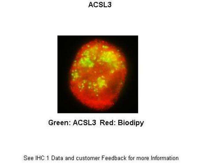
- Immunofluorescence analysis of THP-1 derived macrophage cells using an anti-ACSL3 polyclonal antibody (Product # PA5-42883).
- Submitted by
- Invitrogen Antibodies (provider)
- Main image
- Experimental details
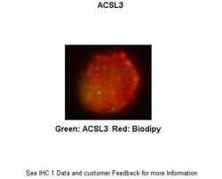
- Immunofluorescence analysis of THP-1 derived macrophage cells using an anti-ACSL3 polyclonal antibody (Product # PA5-42883).
- Submitted by
- Invitrogen Antibodies (provider)
- Main image

- Experimental details
- Immunofluorescence analysis of THP-1 derived macrophage cells using an anti-ACSL3 polyclonal antibody (Product # PA5-42883).

- Submitted by
- Invitrogen Antibodies (provider)
- Main image

- Experimental details
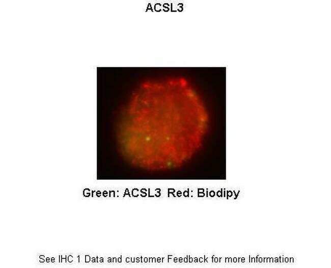
- Immunofluorescence analysis of THP-1 derived macrophage cells using an anti-ACSL3 polyclonal antibody (Product # PA5-42883).

Supportive validation
- Submitted by
- Invitrogen Antibodies (provider)
- Main image
- Experimental details
- NULL
- Submitted by
- Invitrogen Antibodies (provider)
- Main image
- Experimental details
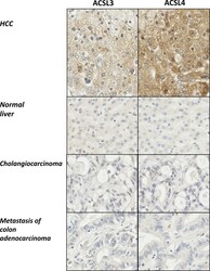
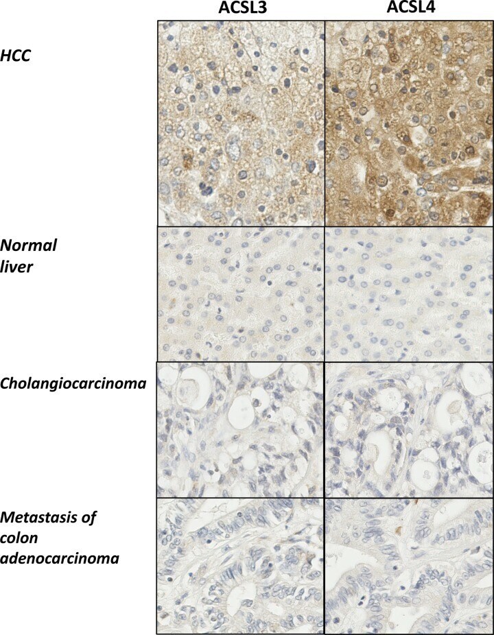
- Figure 1 Immunohistochemistry reveals increased expression of both ACSL3 and ACSL4 in HCC Multiple liver tissues arrays were probed with antibodies specific for either ACSL3 or ACSL4. Representative examples (x20 maginfication) are shown for either ACSL3 or ACSL4 immunohistochemical staining of matched samples of HCC, normal liver, CCA and liver metastases.
- Submitted by
- Invitrogen Antibodies (provider)
- Main image
- Experimental details
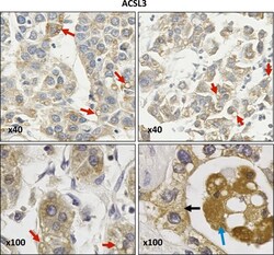
- Figure 3 Anti-ACSL3 IHC staining of HCC samples ACSL3 staining is present on lipid droplets (red arrows) and cytoplasmic reticular membranes (black arrow). Images were obtained at either x40 or x100 magnification.

- Submitted by
- Invitrogen Antibodies (provider)
- Main image
- Experimental details
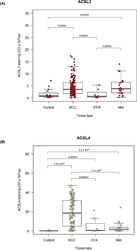
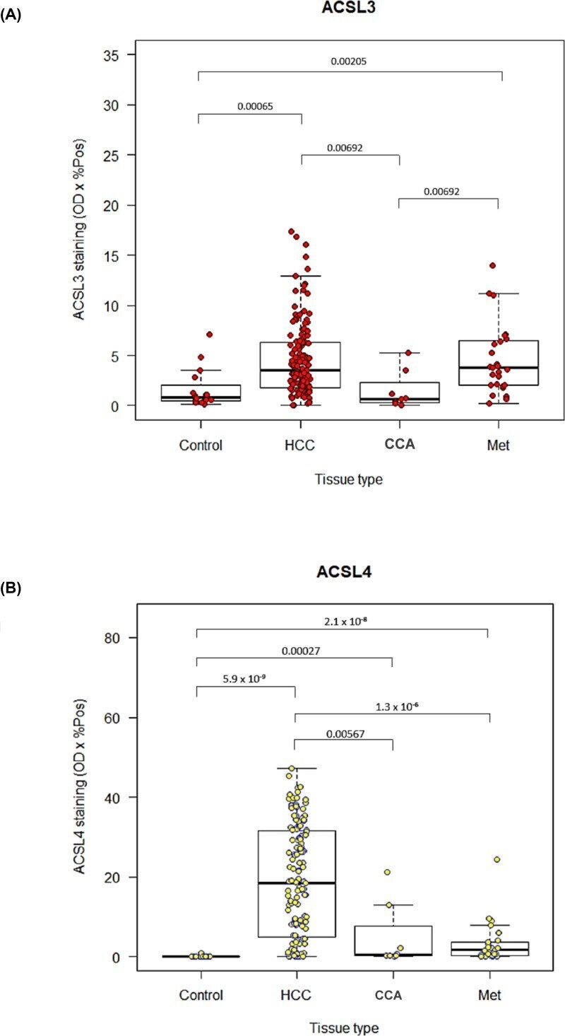
- Figure 4 Expression of ACSL3 and ACSL4 enzymes in hepatic malignancies Computer-aided, quantitative image analysis of ( A ) ACSL3 and ( B ) ACSL4 immunohistochemical staining of a liver tissue microarray for healthy controls, HCC, CCA and hepatic metastases (Met). Box-whisker plots with dot plots overlaid showing the median, interquartile range, minimum and maximum values, ACSL3 and ACSL4 staining values. Pairwise Wilcoxon rank sum tests were performed, significant P -values for which are shown on the graphs.
- Submitted by
- Invitrogen Antibodies (provider)
- Main image
- Experimental details
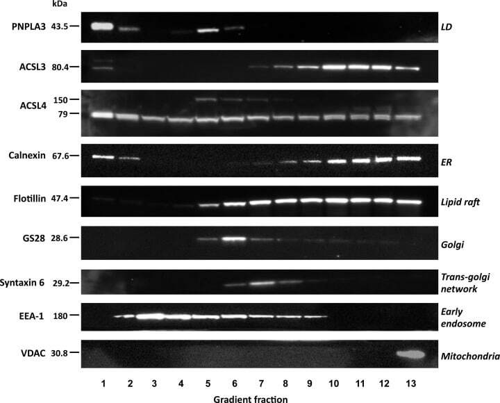
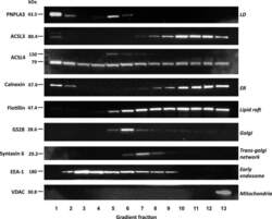
- Figure 6 Equilibrium distributions of ACSL3 and ACSL4 in sucrose density gradient fractions prepared from HepG2 cells Subcellular fractions isolated from HepG2 cells were separated by SDS/PAGE and Western blots were carried out to detect the lipid-droplet protein PNPLA3, ACSL3 and ACSL4, the ER marker protein calnexin, plasma membrane and lipid-raft associated flotillin, the Golgi protein GS28, the TGN-endosomal protein syntaxin-6, the early endosome-recruited protein EEA and the mitochondrial protein VDAC. Western blots are representative of experiments repeated three to four times.