 Explore
 Validate
 Learn
Learn Western blot
 Immunocytochemistry
Antibody data
- Antibody Data
- Antigen structure
- References [1]
- Comments [0]
- Validations
- Immunocytochemistry [2]
- Immunohistochemistry [2]
- Other assay [1]
Submit
Validation data
Reference
Comment
Report error
- Product number
- PA5-29507 - Provider product page

- Provider
- Invitrogen Antibodies
- Product name
- ACSL3 Polyclonal Antibody
- Antibody type
- Polyclonal
- Antigen
- Recombinant full-length protein
- Description
- Recommended positive controls: 293T, A431, Jurkat, Raji. Predicted reactivity: Mouse (95%), Rat (95%), Pig (95%), Bovine (96%). Store product as a concentrated solution. Centrifuge briefly prior to opening the vial.
- Reactivity
- Human
- Host
- Rabbit
- Isotype
- IgG
- Vial size
- 100 μL
- Concentration
- 0.81 mg/mL
- Storage
- Store at 4°C short term. For long term storage, store at -20°C, avoiding freeze/thaw cycles.
Submitted references ACSL3-PAI-1 signaling axis mediates tumor-stroma cross-talk promoting pancreatic cancer progression.
Rossi Sebastiano M, Pozzato C, Saliakoura M, Yang Z, Peng RW, Galiè M, Oberson K, Simon HU, Karamitopoulou E, Konstantinidou G
Science advances 2020 Oct;6(44)
Science advances 2020 Oct;6(44)
No comments: Submit comment
Supportive validation
- Submitted by
- Invitrogen Antibodies (provider)
- Main image

- Experimental details
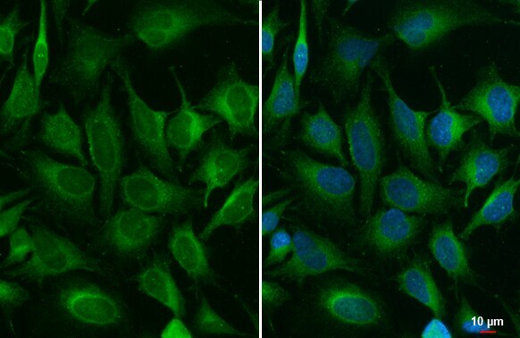
- ACSL3 Polyclonal Antibody detects ACSL3 protein at endoplasmic reticulum by immunofluorescent analysis. Sample: HeLa cells were fixed in 4% paraformaldehyde at RT for 15 min. Green: ACSL3 stained by ACSL3 Polyclonal Antibody (Product # PA5-29507) diluted at 1:500.

- Submitted by
- Invitrogen Antibodies (provider)
- Main image

- Experimental details
- ACSL3 Polyclonal Antibody detects ACSL3 protein at endoplasmic reticulum by immunofluorescent analysis. Sample: HeLa cells were fixed in 4% paraformaldehyde at RT for 15 min. Green: ACSL3 stained by ACSL3 Polyclonal Antibody (Product # PA5-29507) diluted at 1:500.
Supportive validation
- Submitted by
- Invitrogen Antibodies (provider)
- Main image
- Experimental details
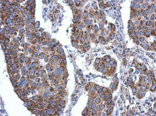
- Immunohistochemical analysis of paraffin-embedded human breast cancer, using ACSL3 (Product # PA5-29507) antibody at 1:500 dilution. Antigen Retrieval: EDTA based buffer, pH 8.0, 15 min.
- Submitted by
- Invitrogen Antibodies (provider)
- Main image
- Experimental details
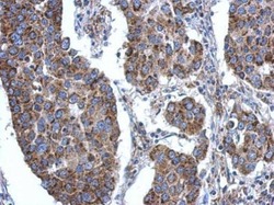
- ACSL3 Polyclonal Antibody detects ACSL3 protein at cytoplasm by immunohistochemical analysis. Sample: Paraffin-embedded human breast carcinoma. ACSL3 stained by ACSL3 Polyclonal Antibody (Product # PA5-29507) diluted at 1:500. Antigen Retrieval: Citrate buffer, pH 6.0, 15 min.

Supportive validation
- Submitted by
- Invitrogen Antibodies (provider)
- Main image
- Experimental details
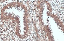
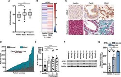
- Fig. 1 ACSL3 is overexpressed in human PDAC. ( A ) mRNA levels of ACSL3 in patient healthy tissue, primary PDAC, and PDAC metastasis from the subset GSE71729. Healthy pancreatic tissue: n = 46 patients, primary PDAC: n = 145 patients, and PDAC metastasis: n = 61 patients. Data are represented as box (Whisker's) plot. Dots evidence outliers according to Tukey's method. ( B ) mRNA levels of ACSL3 in patient primary PDAC and matched adjacent healthy tissue from the subset GSE62452 ( n = 60). Data are expressed as entire numbers normalized by global average. ( C ) Representative IHC staining for ACSL3 (top) and extracellular matrix deposition marker with Masson trichrome (blue, bottom) of a human TMA showing primary healthy tissue, PanIN, and primary PDAC. Scale bars, 50 mum. ( D ) ACSL3 IHC quantification (H-score) of TMA comparing PDAC and adjacent healthy tissue ( n = 50 samples). We considered an H-score of less than 100, from 100 to 200, and above 201 having a low, intermediate, or high staining intensity, respectively. ( E ) Stratification of the patients by tumor grade from (D). Healthy tissue, n = 50 samples; grade I, n = 5 samples; grade II, n = 23 samples; grade III, n = 22 samples. ( F ) Immunoblot for ACSL3, total RAS, and glyceraldehyde-3-phosphate dehydrogenase (GAPDH) of five human patient-derived tumor samples and matched adjacent healthy tissue. ( G ) Correlation between ACSL3 and Masson trichrome staining of TMA from (C) and (D). Samples are divided in low, inte
