Explore
 Validate
 Learn
Learn Western blot
Antibody data
- Antibody Data
- Antigen structure
- References [1]
- Comments [0]
- Validations
- Western blot [3]
- Immunocytochemistry [1]
- Immunohistochemistry [2]
- Other assay [1]
Submit
Validation data
Reference
Comment
Report error
- Product number
- PA5-31227 - Provider product page

- Provider
- Invitrogen Antibodies
- Product name
- GTPBP9 Polyclonal Antibody
- Antibody type
- Polyclonal
- Antigen
- Recombinant full-length protein
- Description
- Recommended positive controls: 293T, A431, HeLa, HepG2, mouse testis, rat testis. Predicted reactivity: Mouse (99%), Rat (99%), Zebrafish (89%), Xenopus laevis (85%), Chicken (90%), Rhesus Monkey (100%), Bovine (98%). Store product as a concentrated solution. Centrifuge briefly prior to opening the vial.
- Reactivity
- Human, Mouse, Rat
- Host
- Rabbit
- Isotype
- IgG
- Vial size
- 100 μL
- Concentration
- 0.46 mg/mL
- Storage
- Store at 4°C short term. For long term storage, store at -20°C, avoiding freeze/thaw cycles.
Submitted references Obg-Like ATPase 1 Enhances Chemoresistance of Breast Cancer via Activation of TGF-β/Smad Axis Cascades.
Liu J, Miao X, Xiao B, Huang J, Tao X, Zhang J, Zhao H, Pan Y, Wang H, Gao G, Xiao GG
Frontiers in pharmacology 2020;11:666
Frontiers in pharmacology 2020;11:666
No comments: Submit comment
Supportive validation
- Submitted by
- Invitrogen Antibodies (provider)
- Main image
- Experimental details
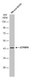
- Western blot analysis of GTPBP9 was performed by separating 50 µg of mouse tissue extract by 10% SDS-PAGE. Proteins were transferred to a membrane and probed with a GTPBP9 Polyclonal Antibody (Product # PA5-31227) at a dilution of 1:1000. The HRP-conjugated anti-rabbit IgG antibody was used to detect the primary antibody.
- Submitted by
- Invitrogen Antibodies (provider)
- Main image
- Experimental details
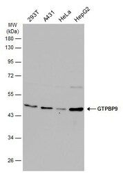
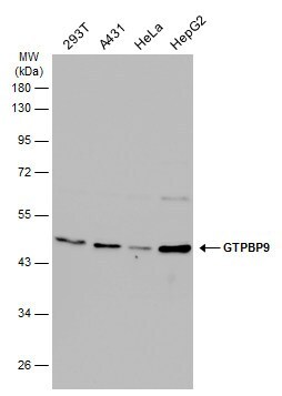
- Western Blot analysis of GTPBP9 was performed by separating 30 µg of various whole cell extracts by 10% SDS-PAGE. Proteins were transferred to a membrane and probed with a GTPBP9 Polyclonal Antibody (Product # PA5-31227) at a dilution of 1:1000 and a HRP-conjugated anti-rabbit IgG secondary antibody.
- Submitted by
- Invitrogen Antibodies (provider)
- Main image
- Experimental details
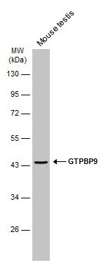
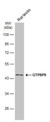
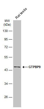
- Western blot analysis of GTPBP9 was performed by separating 50 µg of rat tissue extract by 10% SDS-PAGE. Proteins were transferred to a membrane and probed with a GTPBP9 Polyclonal Antibody (Product # PA5-31227) at a dilution of 1:1000. The HRP-conjugated anti-rabbit IgG antibody was used to detect the primary antibody.
Supportive validation
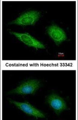
- Submitted by
- Invitrogen Antibodies (provider)
- Main image
- Experimental details
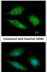
- Immunofluorescent analysis of GTPBP9 in methanol-fixed HeLa cells using a GTPBP9 polyclonal antibody (Product # PA5-31227) at a 1:200 dilution.
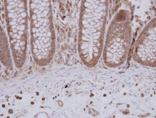
Supportive validation
- Submitted by
- Invitrogen Antibodies (provider)
- Main image

- Experimental details
- Immunohistochemical analysis of paraffin-embedded human colon carcinoma, using GTPBP9 (Product # PA5-31227) antibody at 1:250 dilution. Antigen Retrieval: EDTA based buffer, pH 8.0, 15 min.
- Submitted by
- Invitrogen Antibodies (provider)
- Main image
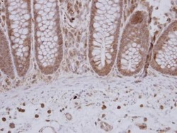
- Experimental details
- Immunohistochemical analysis of paraffin-embedded human colon carcinoma, using GTPBP9 (Product # PA5-31227) antibody at 1:250 dilution. Antigen Retrieval: EDTA based buffer, pH 8.0, 15 min.

Supportive validation
- Submitted by
- Invitrogen Antibodies (provider)
- Main image
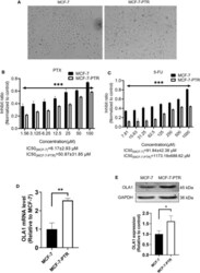
- Experimental details
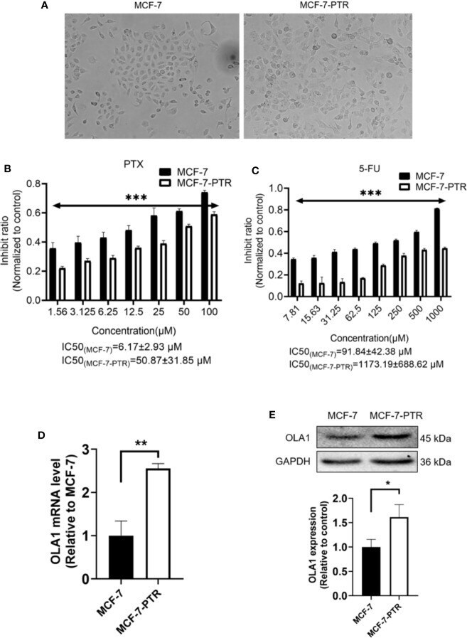
- Figure 2 Upregulated of OLA1 in acquired drug-resistant cell line MCF-7-PTR. (A) Morphology of paclitaxel-induced MCF-7-PTR cells and the parent MCF-7 cells (100X). (B) Drug resistance assay for enhanced expression of OLA1 promotes MCF-7-PTR cell resistance to PTX. (C) Drug resistance assay for enhanced expression of OLA1 promotes MCF-7-PTR cell resistance to 5-Fu. MCF-7 cells and MCF-7-PTR cells were analyzed for the presence of OLA1 by RT-PCR (D) , Western blotting (E) . The relative fold-change was compared with MCF-7 cells (* P < 0.05, ** P < 0.01, *** P < 0.001, Student's t-test).