Explore
 Validate
 Learn
Learn Western blot
 ELISA
ELISA Immunohistochemistry
Antibody data
- Antibody Data
- Antigen structure
- References [3]
- Comments [0]
- Validations
- Immunohistochemistry [4]
- Other assay [1]
Submit
Validation data
Reference
Comment
Report error
- Product number
- PA5-79720 - Provider product page

- Provider
- Invitrogen Antibodies
- Product name
- NKp46 (CD335) Polyclonal Antibody
- Antibody type
- Polyclonal
- Antigen
- Recombinant full-length protein
- Description
- Reconstitute with 0.2 mL of distilled water to yield a concentration of 500 µg/mL. Positive Control - WB: rat spleen tissue, mouse spleen tissue. IHC: rat spleen tissue, mouse spleen tissue, mouse lung tissue, human tonsil tissue.
- Reactivity
- Human, Mouse, Rat
- Host
- Rabbit
- Isotype
- IgG
- Vial size
- 100 μg
- Concentration
- 500 μg/mL
- Storage
- -20°C
Submitted references MITF induces escape from innate immunity in melanoma.
Topographical Distribution and Spatial Interactions of Innate and Semi-Innate Immune Cells in Pancreatic and Other Periampullary Adenocarcinoma.
Liposomal Delivery of Mitoxantrone and a Cholesteryl Indoximod Prodrug Provides Effective Chemo-immunotherapy in Multiple Solid Tumors.
Sánchez-Del-Campo L, Martí-Díaz R, Montenegro MF, González-Guerrero R, Hernández-Caselles T, Martínez-Barba E, Piñero-Madrona A, Cabezas-Herrera J, Goding CR, Rodríguez-López JN
Journal of experimental & clinical cancer research : CR 2021 Mar 31;40(1):117
Journal of experimental & clinical cancer research : CR 2021 Mar 31;40(1):117
Topographical Distribution and Spatial Interactions of Innate and Semi-Innate Immune Cells in Pancreatic and Other Periampullary Adenocarcinoma.
Lundgren S, Micke P, Elebro J, Heby M, Hrynchyk I, Nodin B, Leandersson K, Mezheyeuski A, Jirström K
Frontiers in immunology 2020;11:558169
Frontiers in immunology 2020;11:558169
Liposomal Delivery of Mitoxantrone and a Cholesteryl Indoximod Prodrug Provides Effective Chemo-immunotherapy in Multiple Solid Tumors.
Mei KC, Liao YP, Jiang J, Chiang M, Khazaieli M, Liu X, Wang X, Liu Q, Chang CH, Zhang X, Li J, Ji Y, Melano B, Telesca D, Xia T, Meng H, Nel AE
ACS nano 2020 Oct 27;14(10):13343-13366
ACS nano 2020 Oct 27;14(10):13343-13366
No comments: Submit comment
Supportive validation
- Submitted by
- Invitrogen Antibodies (provider)
- Main image
- Experimental details
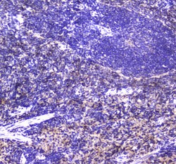
- Immunohistochemistry analysis of NKp46 (CD335) on paraffin-embedded rat spleen tissue. Antigen retrieval was performed using citrate buffer (pH6, epitope retrieval solution) for 20 mins. Sample was blocked using 10% goat serum, incubated with NKp46 (CD335) polyclonal antibody (Product# PA5-79720) with a dilution of 1 µg/mL (overnight at 4°C), and followed by biotinylated goat anti-rabbit IgG (30 minutes at 37°C). Development was performed using Streptavidin-Biotin-Complex (SABC) with DAB chromogen method.

- Submitted by
- Invitrogen Antibodies (provider)
- Main image
- Experimental details
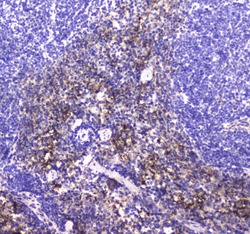
- Immunohistochemistry analysis of NKp46 (CD335) on paraffin-embedded mouse spleen tissue. Antigen retrieval was performed using citrate buffer (pH6, epitope retrieval solution) for 20 mins. Sample was blocked using 10% goat serum, incubated with NKp46 (CD335) polyclonal antibody (Product# PA5-79720) with a dilution of 1 µg/mL (overnight at 4°C), and followed by biotinylated goat anti-rabbit IgG (30 minutes at 37°C). Development was performed using Streptavidin-Biotin-Complex (SABC) with DAB chromogen method.
- Submitted by
- Invitrogen Antibodies (provider)
- Main image
- Experimental details
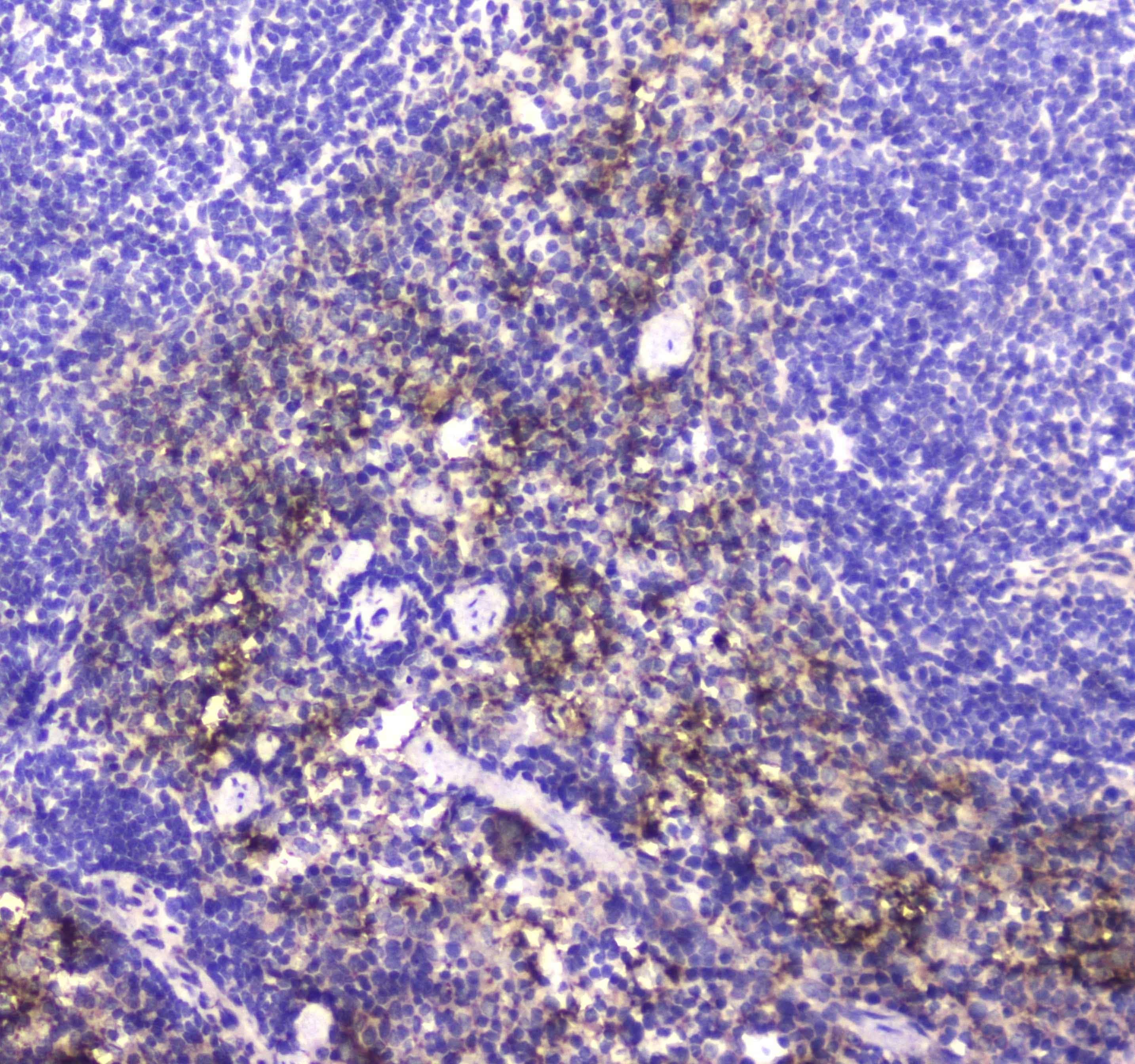
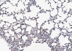
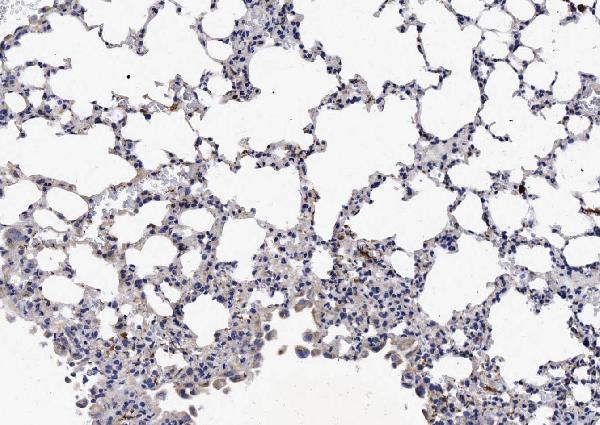
- Immunohistochemical analysis of NCR1 in paraffin-embedded section of mouse lung tissues. Heat mediated antigen retrieval was performed in citrate buffer (pH6, epitope retrieval solution) for 20 mins. The tissue section was blocked with 10% goat serum. The tissue section was then incubated with 1μg/mL rabbit anti-NCR1 antibody (Product # PA5-79720) overnight at 4°C. Biotinylated goat anti-rabbit IgG was used as secondary antibody and incubated for 30 minutes at 37°C. The tissue section was developed using Strepavidin-Biotin-Complex (SABC) with DAB as the chromogen.
- Submitted by
- Invitrogen Antibodies (provider)
- Main image
- Experimental details
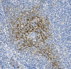
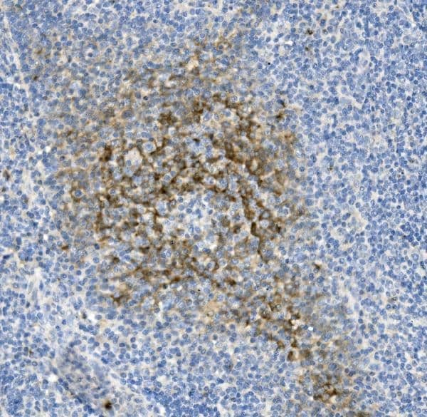
- Immunohistochemical analysis of NCR1 in paraffin-embedded section of human tonsil tissue. Heat mediated antigen retrieval was performed in EDTA buffer (pH 8.0, epitope retrieval solution).The tissue section was blocked with 10% goat serum. The tissue section was then incubated with 1μg/mL rabbit anti-NCR1 antibody (Product # PA5-79720) overnight at 4°C. Biotinylated goat anti-rabbit IgG was used as secondary antibody and incubated for 30 minutes at 37°C. The tissue section was developed using Strepavidin-Biotin-Complex (SABC) with DAB as the chromogen.
Supportive validation
- Submitted by
- Invitrogen Antibodies (provider)
- Main image
- Experimental details
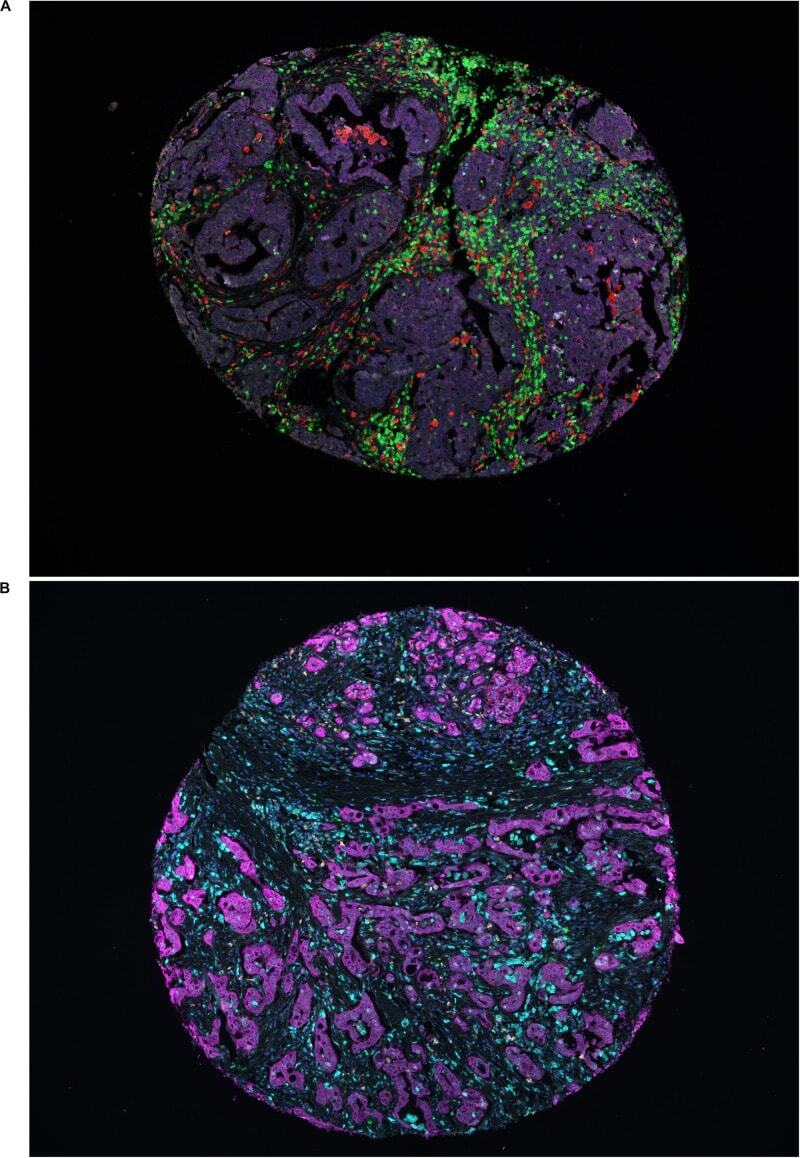
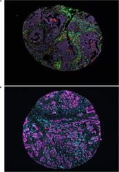
- FIGURE 1 Representative immunofluorescence images. (A) Representative image of a TMA core with heterogeneous infiltration of leukocytes in panel 1; red denoting CD68, yellow denoting CD56, green denoting CD3, pink denoting NKp46, cyan denoting CD163 and magenta denoting cytokeratin. (B) Representative image of a TMA core with heterogeneous infiltration of leukocytes in panel 2; green denoting CD1a, red denoting CD208, yellow denoting CD15, pink denoting CD123, cyan denoting CD68 and magenta denoting cytokeratin.