Explore
 Validate
 Learn
Learn Western blot
 Immunohistochemistry
Antibody data
- Antibody Data
- Antigen structure
- References [1]
- Comments [0]
- Validations
- Western blot [2]
- Immunocytochemistry [1]
- Other assay [1]
Submit
Validation data
Reference
Comment
Report error
- Product number
- PA5-102860 - Provider product page

- Provider
- Invitrogen Antibodies
- Product name
- NKp46 Polyclonal Antibody
- Antibody type
- Polyclonal
- Antigen
- Synthetic peptide
- Description
- Antibody detects endogenous levels of total NCR1.
- Reactivity
- Human, Mouse, Rat
- Host
- Rabbit
- Isotype
- IgG
- Vial size
- 100 μL
- Concentration
- 1 mg/mL
- Storage
- -20°C
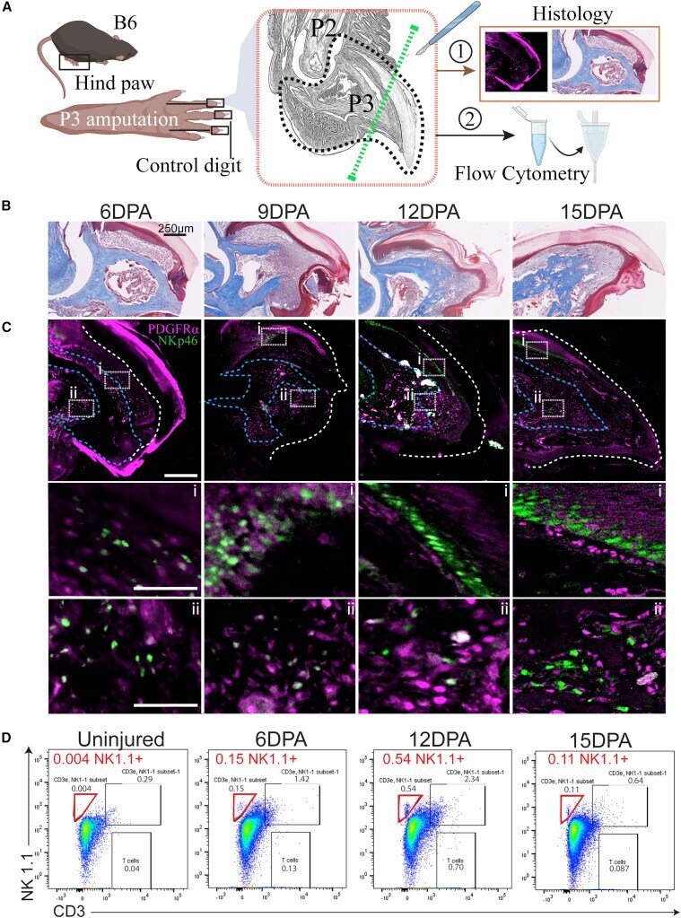
Submitted references Tissue origin of cytotoxic natural killer cells dictates their differential roles in mouse digit tip regeneration and progenitor cell survival.
Dastagir N, Beal Z, Godwin J
Stem cell reports 2022 Mar 8;17(3):633-648
Stem cell reports 2022 Mar 8;17(3):633-648
No comments: Submit comment
Supportive validation
- Submitted by
- Invitrogen Antibodies (provider)
- Main image
- Experimental details
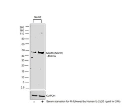
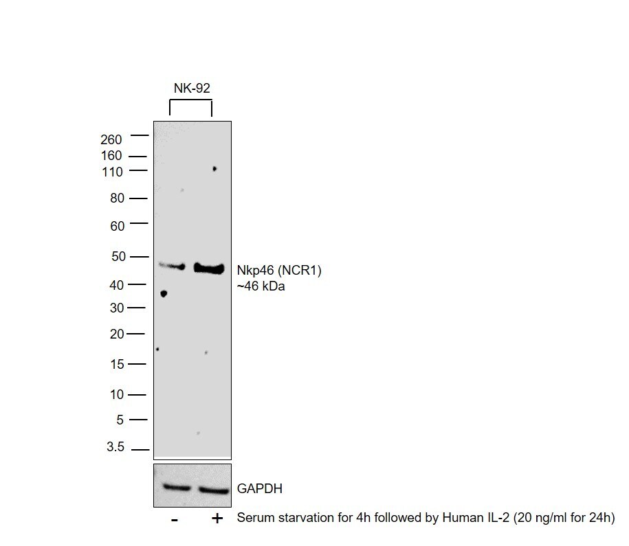
- Western blot was performed using Anti-NKp46 Polyclonal Antibody (Product # PA5-102860) and a 46 kDa band corresponding to Natural cytotoxicity triggering receptor 1 was observed in NK-92 cells which was upregulated upon serum starvation followed by Human IL-2 treatment. Membrane enriched extracts (30 µg lysate) of NK-92 (Lane 1), NK-92 treated with Human IL-2 (20 ng/mL for 24h) (Lane 2) were electrophoresed using NuPAGE™ 4-12% Bis-Tris Protein Gel (Product # NP0322BOX). Resolved proteins were then transferred onto a nitrocellulose membrane (Product # IB23001) by iBlot® 2 Dry Blotting System (Product # IB21001). The blot was probed with the primary antibody (1:1000 dilution) and detected by chemiluminescence with Goat anti-Rabbit IgG (Heavy Chain) Superclonal™ Recombinant Secondary Antibody, HRP (Product # A27036,1:20000 dilution) using the iBright FL 1500 (Product # A32752). Chemiluminescent detection was performed using SuperSignal™ West Pico PLUS Chemiluminescent Substrate (Product # 34580).
- Submitted by
- Invitrogen Antibodies (provider)
- Main image

- Experimental details
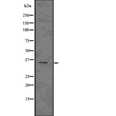
- Western blot analysis of NKp46 in COLO205 cells lysate. Samples were incubated with NKp46 polyclonal antibody (Product # PA5-102860).
Supportive validation
- Submitted by
- Invitrogen Antibodies (provider)
- Main image
- Experimental details
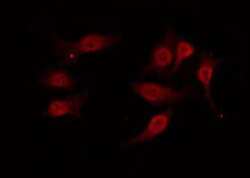
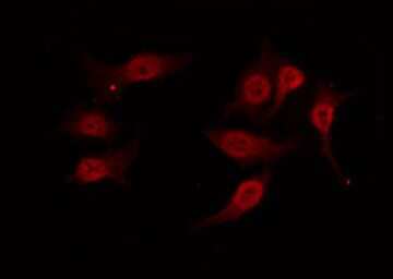
- Immunofluorescent analysis of NKp46 in 293 cells. Samples were fixed with paraformaldehyde, permeabilized with 0.1% Triton X-100, blocked with 10% serum (45 min at 25°C) incubated with NKp46 polyclonal antibody (Product # PA5-102860) using a dilution of 1:200 (1 hr, 37°C), and followed by goat anti-rabbit IgG Alexa Fluor 594 at a dilution of 1:600.
Supportive validation
- Submitted by
- Invitrogen Antibodies (provider)
- Main image
- Experimental details
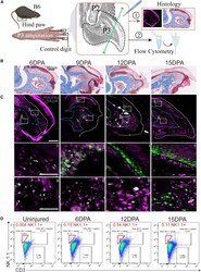
- Figure 1 P3-level digit tip amputations on C57Bl6/J mice show migration of natural killer (NK) cells to the wound site (A) Histological depiction of mouse P2 and P3 bones. The surgery site is indicated by a green dashed line. Tissue taken for FC and histology is indicated by a black dashed line. (B) Early time course of digit tip regeneration using trichrome staining. (C) Representative confocal microscopy with dual staining of PDGFRalpha + (MSCs) and NKP46 + (NK cell) invasion at each time point. (i) and (ii) show high magnification insets. Samples were counterstained with DAPI. White and blue dashed lines mark the nail and soft tissue and bone boundaries, respectively. n = 3; scale bars, 250 mum. (D) Quantification of NK cell recruitment to the wound via FC. NK cells (red gate) defined as CD3-negative and NK1.1-positive peak at 12 DPA and subsequently decrease by 15 DPA. n = 20 digits, five mice/condition. DPA, days post-amputation