Explore
 Validate
 Learn
Learn Western blot
 Immunocytochemistry
Antibody data
- Antibody Data
- Antigen structure
- References [6]
- Comments [0]
- Validations
- Immunocytochemistry [1]
- Immunohistochemistry [1]
Submit
Validation data
Reference
Comment
Report error
- Product number
- AF1850 - Provider product page

- Provider
- R&D Systems
- Product name
- Human NKp46/NCR1 Antibody
- Antibody type
- Polyclonal
- Description
- Antigen Affinity-purified. Detects human NKp46 in direct ELISAs and Western blots. In direct ELISAs, less than 5% cross-reactivity with recombinant mouse NKp46 (MAR-1) is observed and less than 1% cross-reactivity with recombinant human (rh) NKp30, rhNKp44, and rhNKp80 is observed.
- Reactivity
- Human
- Host
- Goat
- Conjugate
- Unconjugated
- Antigen sequence
AAH64806- Isotype
- IgG
- Vial size
- 100 ug
- Concentration
- LYOPH
- Storage
- Use a manual defrost freezer and avoid repeated freeze-thaw cycles. 12 months from date of receipt, -20 to -70 °C as supplied. 1 month, 2 to 8 °C under sterile conditions after reconstitution. 6 months, -20 to -70 °C under sterile conditions after reconstitution.
Submitted references Dipeptidyl Peptidase 4 Inhibitors Reduce Hepatocellular Carcinoma by Activating Lymphocyte Chemotaxis in Mice.
Fc receptor-mediated phagocytosis in tissues as a potent mechanism for preventive and therapeutic HIV vaccine strategies.
Evidence of innate lymphoid cell redundancy in humans.
In situ characterization of intrahepatic non-parenchymal cells in PSC reveals phenotypic patterns associated with disease severity.
Up-regulation of a death receptor renders antiviral T cells susceptible to NK cell-mediated deletion.
Innate immunity in multiple sclerosis white matter lesions: expression of natural cytotoxicity triggering receptor 1 (NCR1).
Nishina S, Yamauchi A, Kawaguchi T, Kaku K, Goto M, Sasaki K, Hara Y, Tomiyama Y, Kuribayashi F, Torimura T, Hino K
Cellular and molecular gastroenterology and hepatology 2019;7(1):115-134
Cellular and molecular gastroenterology and hepatology 2019;7(1):115-134
Fc receptor-mediated phagocytosis in tissues as a potent mechanism for preventive and therapeutic HIV vaccine strategies.
Sips M, Krykbaeva M, Diefenbach TJ, Ghebremichael M, Bowman BA, Dugast AS, Boesch AW, Streeck H, Kwon DS, Ackerman ME, Suscovich TJ, Brouckaert P, Schacker TW, Alter G
Mucosal immunology 2016 Nov;9(6):1584-1595
Mucosal immunology 2016 Nov;9(6):1584-1595
Evidence of innate lymphoid cell redundancy in humans.
Vély F, Barlogis V, Vallentin B, Neven B, Piperoglou C, Ebbo M, Perchet T, Petit M, Yessaad N, Touzot F, Bruneau J, Mahlaoui N, Zucchini N, Farnarier C, Michel G, Moshous D, Blanche S, Dujardin A, Spits H, Distler JH, Ramming A, Picard C, Golub R, Fischer A, Vivier E
Nature immunology 2016 Nov;17(11):1291-1299
Nature immunology 2016 Nov;17(11):1291-1299
In situ characterization of intrahepatic non-parenchymal cells in PSC reveals phenotypic patterns associated with disease severity.
Berglin L, Bergquist A, Johansson H, Glaumann H, Jorns C, Lunemann S, Wedemeyer H, Ellis EC, Björkström NK
PloS one 2014;9(8):e105375
PloS one 2014;9(8):e105375
Up-regulation of a death receptor renders antiviral T cells susceptible to NK cell-mediated deletion.
Peppa D, Gill US, Reynolds G, Easom NJ, Pallett LJ, Schurich A, Micco L, Nebbia G, Singh HD, Adams DH, Kennedy PT, Maini MK
The Journal of experimental medicine 2013 Jan 14;210(1):99-114
The Journal of experimental medicine 2013 Jan 14;210(1):99-114
Innate immunity in multiple sclerosis white matter lesions: expression of natural cytotoxicity triggering receptor 1 (NCR1).
Durrenberger PF, Ettorre A, Kamel F, Webb LV, Sim M, Nicholas RS, Malik O, Reynolds R, Boyton RJ, Altmann DM
Journal of neuroinflammation 2012 Jan 2;9:1
Journal of neuroinflammation 2012 Jan 2;9:1
No comments: Submit comment
Supportive validation
- Submitted by
- R&D Systems (provider)
- Main image

- Experimental details
- NKp46/NCR1 in NK-92 human cell line NKp46/NCR1 was detected in immersion fixed NK-92 human natural killer lymphoma cell line using Goat Anti-Human NKp46/NCR1 Antigen Affinity-purified Polyclonal Antibody (Catalog # AF1850) at 10 µg/mL for 3 hours at room temperature. Cells were stained using the NorthernLights™ 557-conjugated Anti-Goat IgG Secondary Antibody (red; Catalog # NL001) and counterstained with DAPI (blue). View our protocol for Fluorescent ICC Staining of Non-adherent Cells.

Supportive validation
- Submitted by
- R&D Systems (provider)
- Main image

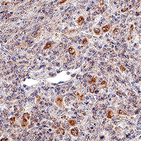
- Experimental details
- NKp46/NCR1 in Human Lymph Node. NKp46/NCR1 was detected in immersion fixed paraffin-embedded sections of human lymph node using Goat Anti-Human NKp46/NCR1 Antigen Affinity-purified Polyclonal Antibody (Catalog # AF1850) at 3 µg/mL for 1 hour at room temperature followed by incubation with the Anti-Goat IgG VisUCyte™ HRP Polymer Antibody (Catalog # VC004). Tissue was stained using DAB (brown) and counterstained with hematoxylin (blue). Specific staining was localized to cytoplasm. View our protocol for IHC Staining with VisUCyte HRP Polymer Detection Reagents.