Explore
 Validate
 Learn
Learn Western blot
 Immunocytochemistry
Antibody data
- Antibody Data
- Antigen structure
- References [1]
- Comments [0]
- Validations
- Immunocytochemistry [2]
- Immunohistochemistry [3]
Submit
Validation data
Reference
Comment
Report error
- Product number
- PA5-27386 - Provider product page

- Provider
- Invitrogen Antibodies
- Product name
- Apolipoprotein D Polyclonal Antibody
- Antibody type
- Polyclonal
- Antigen
- Recombinant full-length protein
- Description
- Recommended positive controls: 293T, A431, HeLa, HepG2. Predicted reactivity: Rabbit (81%), Bovine (83%). Store product as a concentrated solution. Centrifuge briefly prior to opening the vial.
- Reactivity
- Human, Mouse
- Host
- Rabbit
- Isotype
- IgG
- Vial size
- 100 μL
- Concentration
- 1.45 mg/mL
- Storage
- Store at 4°C short term. For long term storage, store at -20°C, avoiding freeze/thaw cycles.
Submitted references LCAT, ApoD, and ApoA1 Expression and Review of Cholesterol Deposition in the Cornea.
Flores R, Jin X, Chang J, Zhang C, Cogan DG, Schaefer EJ, Kruth HS
Biomolecules 2019 Nov 26;9(12)
Biomolecules 2019 Nov 26;9(12)
No comments: Submit comment
Supportive validation
- Submitted by
- Invitrogen Antibodies (provider)
- Main image
- Experimental details
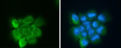
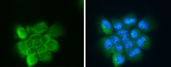
- Apolipoprotein D Polyclonal Antibody detects Apolipoprotein D protein at cytoplasm and nucleus by immunofluorescent analysis. Sample: A431 cells were fixed in 4% paraformaldehyde at RT for 15 min. Green: Apolipoprotein D protein stained by Apolipoprotein D Polyclonal Antibody (Product # PA5-27386) diluted at 1:500. Blue: Hoechst 33342 staining.
- Submitted by
- Invitrogen Antibodies (provider)
- Main image

- Experimental details
- Apolipoprotein D Polyclonal Antibody detects Apolipoprotein D protein at cytoplasm and nucleus by immunofluorescent analysis. Sample: A431 cells were fixed in 4% paraformaldehyde at RT for 15 min. Green: Apolipoprotein D protein stained by Apolipoprotein D Polyclonal Antibody (Product # PA5-27386) diluted at 1:500. Blue: Hoechst 33342 staining.

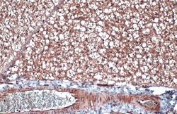
Supportive validation
- Submitted by
- Invitrogen Antibodies (provider)
- Main image

- Experimental details
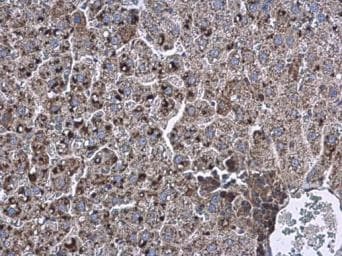
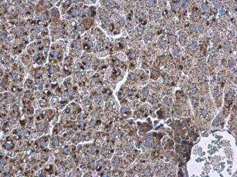
- Immunohistochemistry (Paraffin) analysis of Apolipoprotein D was performed in paraffin-embedded rat liver tissue using Apolipoprotein D Polyclonal Antibody (Product # PA5-27386) at a dilution of 1:500.
- Submitted by
- Invitrogen Antibodies (provider)
- Main image
- Experimental details
- Immunohistochemical analysis of paraffin-embedded mouse brown adipocyte using Apolipoprotein D antibody (Product # PA5-27386) diluted 1:500. Apolipoprotein D protein is detected in the cytoplasm. Citrate buffer (pH 6.0 for 15 minutes) was used for antigen retrieval.

- Submitted by
- Invitrogen Antibodies (provider)
- Main image

- Experimental details
- Immunohistochemical analysis of paraffin-embedded rat liver using Apolipoprotein D antibody (Product # PA5-27386) diluted 1:500. Apolipoprotein D protein is detected in the cytoplasm. Citrate buffer (pH 6.0 for 15 minutes) was used for antigen retrieval.