Explore
 Validate
 Learn
LearnPA5-52665
antibody from Invitrogen Antibodies
Targeting: LAMA3
BM600-150kDa, epiligrin, kalinin-165kDa, LAMNA, nicein-150kDa
 Immunocytochemistry
Antibody data
- Antibody Data
- Antigen structure
- References [1]
- Comments [0]
- Validations
- Immunocytochemistry [2]
- Immunohistochemistry [1]
- Other assay [1]
Submit
Validation data
Reference
Comment
Report error
- Product number
- PA5-52665 - Provider product page

- Provider
- Invitrogen Antibodies
- Product name
- Laminin alpha-3 Polyclonal Antibody
- Antibody type
- Polyclonal
- Antigen
- Recombinant protein fragment
- Description
- Immunogen sequence: LLNRIRTWQK THQGENNGLA NSIRDSLNEY EAKLSDLRAR LQEAAAQAKQ ANGLNQENER ALGAIQRQVK EINSLQSDFT KYLTTADSSL LQTNIALQLM EKSQKEYE Highest antigen sequence identity to the following orthologs: Mouse - 74%, Rat - 71%.
- Reactivity
- Human
- Host
- Rabbit
- Isotype
- IgG
- Vial size
- 100 μL
- Concentration
- 0.3 mg/mL
- Storage
- Store at 4°C short term. For long term storage, store at -20°C, avoiding freeze/thaw cycles.
Submitted references Evolving Up-regulation of Biliary Fibrosis-Related Extracellular Matrix Molecules After Successful Portoenterostomy.
Kyrönlahti A, Godbole N, Akinrinade O, Soini T, Nyholm I, Andersson N, Hukkinen M, Lohi J, Wilson DB, Pihlajoki M, Pakarinen MP, Heikinheimo M
Hepatology communications 2021 Jun;5(6):1036-1050
Hepatology communications 2021 Jun;5(6):1036-1050
No comments: Submit comment
Supportive validation
- Submitted by
- Invitrogen Antibodies (provider)
- Main image

- Experimental details
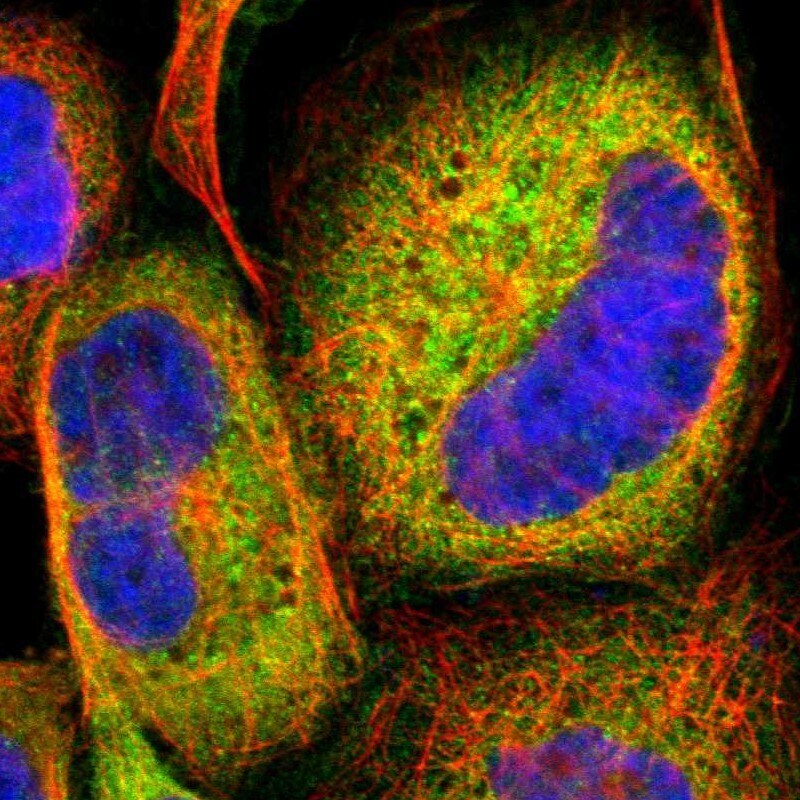
- Immunofluorescent staining of Laminin alpha-3 in human cell line A-431 using Laminin alpha-3 Polyclonal Antibody (Product # PA5-52665) shows localization to endoplasmic reticulum.

- Submitted by
- Invitrogen Antibodies (provider)
- Main image

- Experimental details
- Immunofluorescent staining of Laminin alpha-3 in human cell line A-431 using Laminin alpha-3 Polyclonal Antibody (Product # PA5-52665) shows localization to endoplasmic reticulum.
Supportive validation
- Submitted by
- Invitrogen Antibodies (provider)
- Main image
- Experimental details
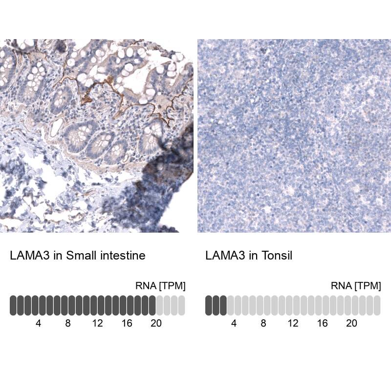
- Immunohistochemical analysis of Laminin alpha-3 in human kidney using Laminin alpha-3 Polyclonal Antibody (Product # PA5-52665) shows strong cytoplasmic positivity in cells in tubules.
Supportive validation
- Submitted by
- Invitrogen Antibodies (provider)
- Main image
- Experimental details
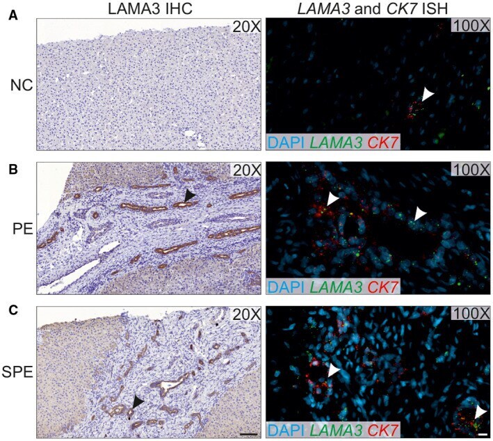
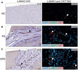
- FIG. 6 Abnormal expression of LAMA3 in neo-ductular areas of BA livers. Liver samples from NC (A), PE (B), and SPE (C) were subjected to ISH or IHC. Expression of LAMA3 protein and LAMA3 as well as CK7 mRNA are shown in the left and right panels, respectively. Increased LAMA3 expression with a consistent co-expression of CK7 in ductular cholangiocytes can be seen within areas of DR in the PE and SPE samples. Brown indicates the positive staining in IHC panels. The black arrowhead indicates increased LAMA3 protein expression, and the white arrowhead indicates the LAMA3 mRNA expression in the ductular cholangiocytes. Bars: IHC, 100 mum; ISH, 10 mum.