 Explore
 Validate
 Learn
LearnMAB21441
antibody from R&D Systems
Targeting: LAMA3
BM600-150kDa, epiligrin, kalinin-165kDa, LAMNA, nicein-150kDa
 Western blot
 Immunoprecipitation
Antibody data
- Antibody Data
- Antigen structure
- References [1]
- Comments [0]
- Validations
- Western blot [1]
- Immunocytochemistry [1]
- Flow cytometry [1]
Submit
Validation data
Reference
Comment
Report error
- Product number
- MAB21441 - Provider product page

- Provider
- R&D Systems
- Product name
- Anti-Human Laminin alpha 3/Laminin-5 Monoclonal Antibody (Clone 546215)
- Antibody type
- Monoclonal
- Antigen
- Chinese hamster ovary cell line CHO-derived recombinant human Laminin α3/Laminin‑5, aa 21-1713
- Description
- Protein A or G purified from hybridoma culture supernatant
- Reactivity
- Human
- Host
- Mouse
- Antigen sequence
NP_000218- Isotype
- IgG
- Vial size
- 100 µg
Submitted references Laminin signals initiate the reciprocal loop that informs breast-specific gene expression and homeostasis by activating NO, p53 and microRNAs.
Furuta S, Ren G, Mao JH, Bissell MJ
eLife 2018 Mar 21;7
eLife 2018 Mar 21;7
No comments: Submit comment
Supportive validation
- Submitted by
- R&D Systems (provider)
- Main image
- Experimental details
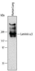
- Detection of Human Laminin alpha 3/Laminin-5 by Western Blot. Western blot shows lysates of human lung tissue. PVDF Membrane was probed with 2 µg/mL of Mouse Anti-Human Laminin alpha 3/ Laminin-5 Monoclonal Antibody (Catalog # MAB21441) followed by HRP-conjugated Anti-Mouse IgG Secondary Antibody (Catalog # HAF007). A specific band was detected for Laminin alpha 3/Laminin-5 at approximately 190 kDa (as indicated). This experiment was conducted under non-reducing conditions and using Immunoblot Buffer Group 1.

Supportive validation
- Submitted by
- R&D Systems (provider)
- Main image
- Experimental details
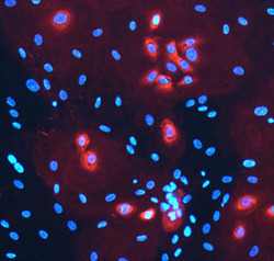
- Laminin alpha 3/Laminin-5 in NHEK Human Cell Line. Laminin alpha 3/Laminin-5 was detected in immersion fixed NHEK human normal epidermal keratinocytes treated with 10ng/mL Recombinant Human TGF-beta 1 (Catalog # 240-B) for 24 hours using Mouse Anti-Human Laminin alpha 3/Laminin-5 Monoclonal Antibody (Catalog # MAB21441) at 10 µg/mL for 3 hours at room temperature. Cells were stained using the NorthernLights™ 557-conjugated Anti-Mouse IgG Secondary Antibody (red; Catalog # NL007) and counterstained with DAPI (blue). Specific staining was localized to the perinuclear space. View our protocol for Fluorescent ICC Staining of Cells on Coverslips.

Supportive validation
- Submitted by
- R&D Systems (provider)
- Main image
- Experimental details
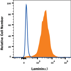
- Detection of Laminin alpha 3/Laminin-5 in U2OS Human Cell Line by Flow Cytometry. U2OS human osteosarcoma cell line was stained with Mouse Anti-Human Laminin alpha 3/Laminin-5 Monoclonal Antibody (Catalog # MAB21441, filled histogram) or isotype control antibody (Catalog # MAB002, open histogram), followed by Allophycocyanin-conjugated Anti-Mouse IgG Secondary Antibody (Catalog # F0101B). View our protocol for Staining Intracellular Molecules.
