 Explore
 Validate
 Learn
Learn Western blot
 Immunocytochemistry
Antibody data
- Antibody Data
- Antigen structure
- References [13]
- Comments [0]
- Validations
- Immunocytochemistry [5]
- Immunohistochemistry [3]
Submit
Validation data
Reference
Comment
Report error
- Product number
- MA3-043 - Provider product page

- Provider
- Invitrogen Antibodies
- Product name
- CHRNA1 Monoclonal Antibody (88B)
- Antibody type
- Monoclonal
- Antigen
- Purifed from natural sources
- Description
- MA3-043 detects nicotinic acetylcholine receptor (nAChR) gamma and delta subunits in torpedo and the delta subunit in mouse, human, rat, chicken and frog. This antibody does not detect the alpha 1 and beta 1 subunits. MA3-043 has been successfully used in Western blot, immunohistochemistry and immunoprecipitation procedures. By Western blot, this antibody detects a 60 kDa protein representing the gamma subunit and a 65 kDa protein representing the delta subunit from torpedo skeletal muscle homogenates. Under non-reducing conditions the delta subunit migrates mostly as a dimer of ~130 kDa. Immunohistochemical staining of nAChR in rat skeletal muscle with MA3-043 results in strong staining of the motor endplate. In Immunohistochemical staining, cryostat or permeabilization of the sections is recommended due to the reactivity of this antibody with the cytoplasmic side of the receptor. The MA3-043 antigen is purified Torpedo californica acetylcholine receptor.
- Reactivity
- Human, Mouse, Rat, Chicken/Avian
- Host
- Mouse
- Isotype
- IgG
- Antibody clone number
- 88B
- Vial size
- 100 μL
- Concentration
- Conc. Not Determined
- Storage
- -20°C, Avoid Freeze/Thaw Cycles
Submitted references RNU (Foxn1 (RNU)-Nude) Rats Demonstrate an Improved Ability to Regenerate Muscle in a Volumetric Muscle Injury Compared to Sprague Dawley Rats.
Expression of the skeletal muscle dystrophin-dystroglycan complex and syntrophin-nitric oxide synthase complex is severely affected in the type 2 diabetic Goto-Kakizaki rat.
Increased expression of the nicotinic acetylcholine receptor in stimulated muscle.
Increased expression of the nicotinic acetylcholine receptor in stimulated muscle.
Use of continuous-elution gel electrophoresis as a preparative tool for blot overlay analysis.
Use of continuous-elution gel electrophoresis as a preparative tool for blot overlay analysis.
Epsilon subunit-containing acetylcholine receptors in myotubes belong to the slowly degrading population.
Association of acetylcholine receptors with peripheral membrane proteins: evidence from antibody-induced coaggregation.
Association of acetylcholine receptors with peripheral membrane proteins: evidence from antibody-induced coaggregation.
Dystrophin in a membrane skeletal network: localization and comparison to other proteins.
Dystrophin in a membrane skeletal network: localization and comparison to other proteins.
Monoclonal antibody identifies a 200-kDa subunit of the dihydropyridine-sensitive calcium channel.
Monoclonal antibodies to cytoplasmic domains of the acetylcholine receptor.
McClure MJ, Olson LC, Cohen DJ, Huang YC, Zhang S, Nguyen T, Boyan BD, Schwartz Z
Bioengineering (Basel, Switzerland) 2021 Jan 15;8(1)
Bioengineering (Basel, Switzerland) 2021 Jan 15;8(1)
Expression of the skeletal muscle dystrophin-dystroglycan complex and syntrophin-nitric oxide synthase complex is severely affected in the type 2 diabetic Goto-Kakizaki rat.
Mulvey C, Harno E, Keenan A, Ohlendieck K
European journal of cell biology 2005 Nov;84(11):867-83
European journal of cell biology 2005 Nov;84(11):867-83
Increased expression of the nicotinic acetylcholine receptor in stimulated muscle.
O'Reilly C, Pette D, Ohlendieck K
Biochemical and biophysical research communications 2003 Jan 10;300(2):585-91
Biochemical and biophysical research communications 2003 Jan 10;300(2):585-91
Increased expression of the nicotinic acetylcholine receptor in stimulated muscle.
O'Reilly C, Pette D, Ohlendieck K
Biochemical and biophysical research communications 2003 Jan 10;300(2):585-91
Biochemical and biophysical research communications 2003 Jan 10;300(2):585-91
Use of continuous-elution gel electrophoresis as a preparative tool for blot overlay analysis.
Mulvey C, Ohlendieck K
Analytical biochemistry 2003 Aug 1;319(1):122-30
Analytical biochemistry 2003 Aug 1;319(1):122-30
Use of continuous-elution gel electrophoresis as a preparative tool for blot overlay analysis.
Mulvey C, Ohlendieck K
Analytical biochemistry 2003 Aug 1;319(1):122-30
Analytical biochemistry 2003 Aug 1;319(1):122-30
Epsilon subunit-containing acetylcholine receptors in myotubes belong to the slowly degrading population.
Sala C, O'Malley J, Xu R, Fumagalli G, Salpeter MM
The Journal of neuroscience : the official journal of the Society for Neuroscience 1997 Dec 1;17(23):8937-44
The Journal of neuroscience : the official journal of the Society for Neuroscience 1997 Dec 1;17(23):8937-44
Association of acetylcholine receptors with peripheral membrane proteins: evidence from antibody-induced coaggregation.
Bloch RJ, Sealock R, Pumplin DW, Luther PW, Froehner SC
The Journal of membrane biology 1994 Feb;138(1):13-28
The Journal of membrane biology 1994 Feb;138(1):13-28
Association of acetylcholine receptors with peripheral membrane proteins: evidence from antibody-induced coaggregation.
Bloch RJ, Sealock R, Pumplin DW, Luther PW, Froehner SC
The Journal of membrane biology 1994 Feb;138(1):13-28
The Journal of membrane biology 1994 Feb;138(1):13-28
Dystrophin in a membrane skeletal network: localization and comparison to other proteins.
Dmytrenko GM, Pumplin DW, Bloch RJ
The Journal of neuroscience : the official journal of the Society for Neuroscience 1993 Feb;13(2):547-58
The Journal of neuroscience : the official journal of the Society for Neuroscience 1993 Feb;13(2):547-58
Monoclonal antibody identifies a 200-kDa subunit of the dihydropyridine-sensitive calcium channel.
Morton ME, Froehner SC
The Journal of biological chemistry 1987 Sep 5;262(25):11904-7
The Journal of biological chemistry 1987 Sep 5;262(25):11904-7
Monoclonal antibodies to cytoplasmic domains of the acetylcholine receptor.
Froehner SC, Douville K, Klink S, Culp WJ
The Journal of biological chemistry 1983 Jun 10;258(11):7112-20
The Journal of biological chemistry 1983 Jun 10;258(11):7112-20
No comments: Submit comment
Supportive validation
- Submitted by
- Invitrogen Antibodies (provider)
- Main image

- Experimental details
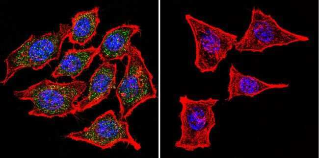
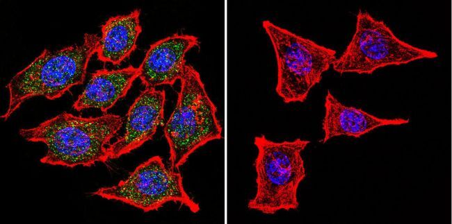
- Immunofluorescent analysis of Nicotinic Acetylcholine Receptor using Anti-Nicotinic Acetylcholine Receptor Monoclonal Antibody (88B) (Product # MA3-043) shows staining in Hela Cells. Nicotinic Acetylcholine Receptor staining (green), F-Actin staining with Phalloidin (red) and nuclei with DAPI (blue) is shown. Cells were grown on chamber slides and fixed with formaldehyde prior to staining. Cells were probed without (control) or with or an antibody recognizing Nicotinic Acetylcholine Receptor (Product # MA3-043) at a dilution of 1:100 over night at 4°C, washed with PBS and incubated with a DyLight-488 conjugated secondary antibody (Product # 35503, Goat Anti-Mouse). Images were taken at 60X magnification.
- Submitted by
- Invitrogen Antibodies (provider)
- Main image
- Experimental details
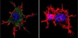
- Immunofluorescent analysis of Nicotinic Acetylcholine Receptor using Anti-Nicotinic Acetylcholine Receptor Monoclonal Antibody (88B) (Product # MA3-043) shows staining in Neuro-2a Cells. Nicotinic Acetylcholine Receptor staining (green), F-Actin staining with Phalloidin (red) and nuclei with DAPI (blue) is shown. Cells were grown on chamber slides and fixed with formaldehyde prior to staining. Cells were probed without (control) or with or an antibody recognizing Nicotinic Acetylcholine Receptor (Product # MA3-043) at a dilution of 1:100 over night at 4°C, washed with PBS and incubated with a DyLight-488 conjugated secondary antibody (Product # 35503, Goat Anti-Mouse). Images were taken at 60X magnification.
- Submitted by
- Invitrogen Antibodies (provider)
- Main image
- Experimental details
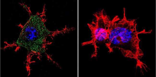
- Immunofluorescent analysis of Nicotinic Acetylcholine Receptor using Anti-Nicotinic Acetylcholine Receptor Monoclonal Antibody (88B) (Product # MA3-043) shows staining in U251 Cells. Nicotinic Acetylcholine Receptor staining (green), F-Actin staining with Phalloidin (red) and nuclei with DAPI (blue) is shown. Cells were grown on chamber slides and fixed with formaldehyde prior to staining. Cells were probed without (control) or with or an antibody recognizing Nicotinic Acetylcholine Receptor (Product # MA3-043) at a dilution of 1:20 over night at 4°C, washed with PBS and incubated with a DyLight-488 conjugated secondary antibody (Product # 35503, Goat Anti-Mouse). Images were taken at 60X magnification.

- Submitted by
- Invitrogen Antibodies (provider)
- Main image

- Experimental details
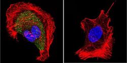
- Immunofluorescent analysis of Nicotinic Acetylcholine Receptor using Anti-Nicotinic Acetylcholine Receptor Monoclonal Antibody (88B) (Product # MA3-043) shows staining in Hela Cells. Nicotinic Acetylcholine Receptor staining (green), F-Actin staining with Phalloidin (red) and nuclei with DAPI (blue) is shown. Cells were grown on chamber slides and fixed with formaldehyde prior to staining. Cells were probed without (control) or with or an antibody recognizing Nicotinic Acetylcholine Receptor (Product # MA3-043) at a dilution of 1:100 over night at 4°C, washed with PBS and incubated with a DyLight-488 conjugated secondary antibody (Product # 35503, Goat Anti-Mouse). Images were taken at 60X magnification.
- Submitted by
- Invitrogen Antibodies (provider)
- Main image

- Experimental details
- Immunofluorescent analysis of Nicotinic Acetylcholine Receptor using Anti-Nicotinic Acetylcholine Receptor Monoclonal Antibody (88B) (Product # MA3-043) shows staining in Neuro-2a Cells. Nicotinic Acetylcholine Receptor staining (green), F-Actin staining with Phalloidin (red) and nuclei with DAPI (blue) is shown. Cells were grown on chamber slides and fixed with formaldehyde prior to staining. Cells were probed without (control) or with or an antibody recognizing Nicotinic Acetylcholine Receptor (Product # MA3-043) at a dilution of 1:100 over night at 4°C, washed with PBS and incubated with a DyLight-488 conjugated secondary antibody (Product # 35503, Goat Anti-Mouse). Images were taken at 60X magnification.

Supportive validation
- Submitted by
- Invitrogen Antibodies (provider)
- Main image
- Experimental details
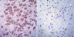
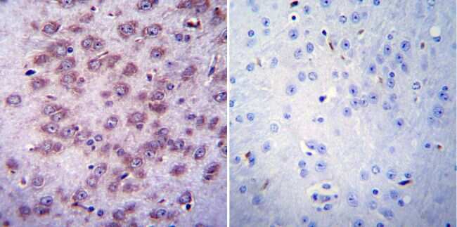
- Immunohistochemistry was performed on normal biopsies of deparaffinized Mouse brain tissue. To expose target proteins, heat induced antigen retrieval was performed using 10mM sodium citrate (pH6.0) buffer, microwaved for 8-15 minutes. Following antigen retrieval tissues were blocked in 3% BSA-PBS for 30 minutes at room temperature. Tissues were then probed at a dilution of 1:20 with a mouse monoclonal antibody recognizing Nicotinic Acetylcholine Receptor (Product # MA3-043) or without primary antibody (negative control) overnight at 4°C in a humidified chamber. Tissues were washed extensively with PBST and endogenous peroxidase activity was quenched with a peroxidase suppressor. Detection was performed using a biotin-conjugated secondary antibody and SA-HRP, followed by colorimetric detection using DAB. Tissues were counterstained with hematoxylin and prepped for mounting.
- Submitted by
- Invitrogen Antibodies (provider)
- Main image
- Experimental details
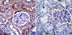
- Immunohistochemistry was performed on normal biopsies of deparaffinized Mouse kidney tissue. To expose target proteins, heat induced antigen retrieval was performed using 10mM sodium citrate (pH6.0) buffer, microwaved for 8-15 minutes. Following antigen retrieval tissues were blocked in 3% BSA-PBS for 30 minutes at room temperature. Tissues were then probed at a dilution of 1:20 with a mouse monoclonal antibody recognizing Nicotinic Acetylcholine Receptor (Product # MA3-043) or without primary antibody (negative control) overnight at 4°C in a humidified chamber. Tissues were washed extensively with PBST and endogenous peroxidase activity was quenched with a peroxidase suppressor. Detection was performed using a biotin-conjugated secondary antibody and SA-HRP, followed by colorimetric detection using DAB. Tissues were counterstained with hematoxylin and prepped for mounting.

- Submitted by
- Invitrogen Antibodies (provider)
- Main image
- Experimental details
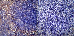
- Immunohistochemistry was performed on normal biopsies of deparaffinized Mouse lymph node tissue. To expose target proteins, heat induced antigen retrieval was performed using 10mM sodium citrate (pH6.0) buffer, microwaved for 8-15 minutes. Following antigen retrieval tissues were blocked in 3% BSA-PBS for 30 minutes at room temperature. Tissues were then probed at a dilution of 1:20 with a mouse monoclonal antibody recognizing Nicotinic Acetylcholine Receptor (Product # MA3-043) or without primary antibody (negative control) overnight at 4°C in a humidified chamber. Tissues were washed extensively with PBST and endogenous peroxidase activity was quenched with a peroxidase suppressor. Detection was performed using a biotin-conjugated secondary antibody and SA-HRP, followed by colorimetric detection using DAB. Tissues were counterstained with hematoxylin and prepped for mounting.
