Explore
 Validate
 Learn
Learn Western blot
 Immunocytochemistry
Antibody data
- Antibody Data
- Antigen structure
- References [4]
- Comments [0]
- Validations
- Immunocytochemistry [1]
- Immunohistochemistry [1]
Submit
Validation data
Reference
Comment
Report error
- Product number
- HPA022015 - Provider product page

- Provider
- Atlas Antibodies
- Proper citation
- Atlas Antibodies Cat#HPA022015, RRID:AB_1846462
- Product name
- Anti-CDR2L
- Antibody type
- Polyclonal
- Description
- Polyclonal Antibody against Human CDR2L, Gene description: cerebellar degeneration-related protein 2-like, Alternative Gene Names: HUMPPA, Validated applications: WB, IHC, ICC, Uniprot ID: Q86X02, Storage: Store at +4°C for short term storage. Long time storage is recommended at -20°C.
- Reactivity
- Human, Mouse, Rat
- Host
- Rabbit
- Conjugate
- Unconjugated
- Isotype
- IgG
- Vial size
- 100 µl
- Concentration
- 0.1 mg/ml
- Storage
- Store at +4°C for short term storage. Long time storage is recommended at -20°C.
- Handling
- The antibody solution should be gently mixed before use.
Submitted references Paraneoplastic cerebellar degeneration: Yo antibody alters mitochondrial calcium buffering capacity
Cerebellar degeneration-related proteins 2 and 2-like are present in ovarian cancer in patients with and without Yo antibodies
Paraneoplastic CDR2 and CDR2L antibodies affect Purkinje cell calcium homeostasis
CDR2L Antibodies: A New Player in Paraneoplastic Cerebellar Degeneration
Panja D, Vedeler C, Schubert M
Neuropathology and Applied Neurobiology 2018;45(2):141-156
Neuropathology and Applied Neurobiology 2018;45(2):141-156
Cerebellar degeneration-related proteins 2 and 2-like are present in ovarian cancer in patients with and without Yo antibodies
Raspotnig M, Haugen M, Thorsteinsdottir M, Stefansson I, Salvesen H, Storstein A, Vedeler C
Cancer Immunology, Immunotherapy 2017;66(11):1463-1471
Cancer Immunology, Immunotherapy 2017;66(11):1463-1471
Paraneoplastic CDR2 and CDR2L antibodies affect Purkinje cell calcium homeostasis
Schubert M, Panja D, Haugen M, Bramham C, Vedeler C
Acta Neuropathologica 2014;128(6):835-852
Acta Neuropathologica 2014;128(6):835-852
CDR2L Antibodies: A New Player in Paraneoplastic Cerebellar Degeneration
Datta P, Eichler T, Totland C, Haugen M, Qvale T, Mazengia K, Storstein A, Haukanes B, Vedeler C
PLoS ONE 2013;8(6):e66002
PLoS ONE 2013;8(6):e66002
No comments: Submit comment
Supportive validation
- Submitted by
- Atlas Antibodies (provider)
- Main image
- Experimental details
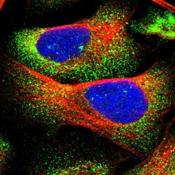
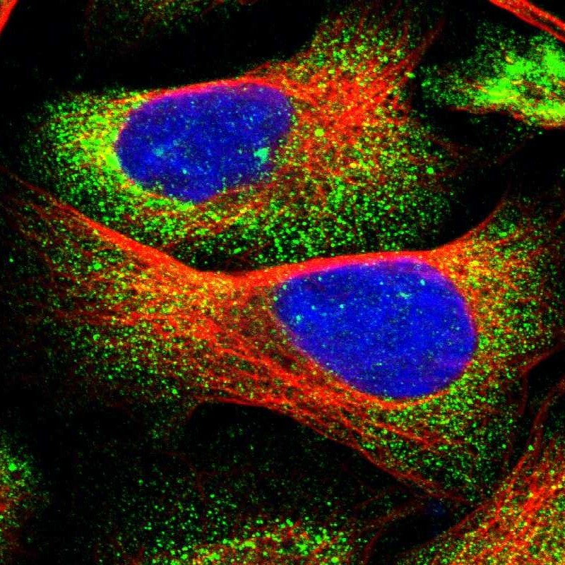
- Immunofluorescent staining of human cell line U-2 OS shows localization to cytosol.
- Sample type
- Human
Supportive validation
- Submitted by
- Atlas Antibodies (provider)
- Enhanced method
- Orthogonal validation
- Main image
- Experimental details
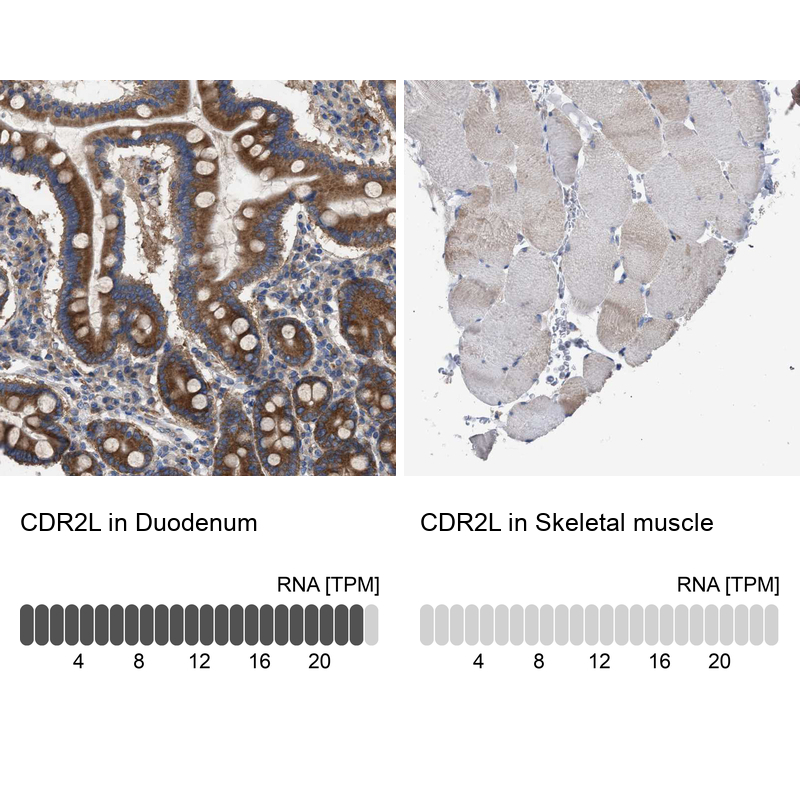
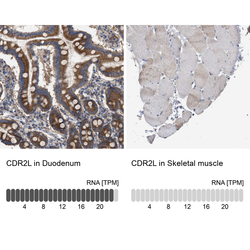
- Immunohistochemistry analysis in human duodenum and skeletal muscle tissues using HPA022015 antibody. Corresponding CDR2L RNA-seq data are presented for the same tissues.
- Sample type
- Human
- Protocol
- Protocol