Explore
 Validate
 Learn
Learn Western blot
 Immunohistochemistry
Antibody data
- Antibody Data
- Antigen structure
- References [3]
- Comments [0]
- Validations
- Immunohistochemistry [1]
Submit
Validation data
Reference
Comment
Report error
- Product number
- HPA028825 - Provider product page

- Provider
- Atlas Antibodies
- Proper citation
- Atlas Antibodies Cat#HPA028825, RRID:AB_10602130
- Product name
- Anti-FABP7
- Antibody type
- Polyclonal
- Description
- Polyclonal Antibody against Human FABP7, Gene description: fatty acid binding protein 7, brain, Alternative Gene Names: B-FABP, BLBP, Validated applications: WB, IHC, Uniprot ID: O15540, Storage: Store at +4°C for short term storage. Long time storage is recommended at -20°C.
- Reactivity
- Human
- Host
- Rabbit
- Conjugate
- Unconjugated
- Isotype
- IgG
- Vial size
- 100 µl
- Concentration
- 0.1 mg/ml
- Storage
- Store at +4°C for short term storage. Long time storage is recommended at -20°C.
- Handling
- The antibody solution should be gently mixed before use.
Submitted references
FABP7 and HMGCS2 are novel protein markers for apocrine differentiation categorizing apocrine carcinoma of the breast.
Impact of Gender in Renal Cell Carcinoma: The Relationship of FABP7 and BRN2 Expression with Overall Survival
Paryani F, Kwon J, Ng C, Madden N, Ofori K, Tang A, Lu H, Li J, Mahajan A, Davidson S, Basile A, McHugh C, Vonsattel J, Hickman R, Zody M, Houseman D, Goldman J, Yoo A, Menon V, Al-Dalahmah O
2023
2023
FABP7 and HMGCS2 are novel protein markers for apocrine differentiation categorizing apocrine carcinoma of the breast.
Gromov P, Espinoza JA, Talman ML, Honma N, Kroman N, Timmermans Wielenga V, Moreira JM, Gromova I
PloS one 2014;9(11):e112024
PloS one 2014;9(11):e112024
Impact of Gender in Renal Cell Carcinoma: The Relationship of FABP7 and BRN2 Expression with Overall Survival
Tan C, Takayama T, Takaoka N, Fujita H, Miyazaki M, Sugiyama T, Ozono S
Clinical Medicine Insights: Oncology 2014;8
Clinical Medicine Insights: Oncology 2014;8
No comments: Submit comment
Supportive validation
- Submitted by
- Atlas Antibodies (provider)
- Enhanced method
- Orthogonal validation
- Main image
- Experimental details
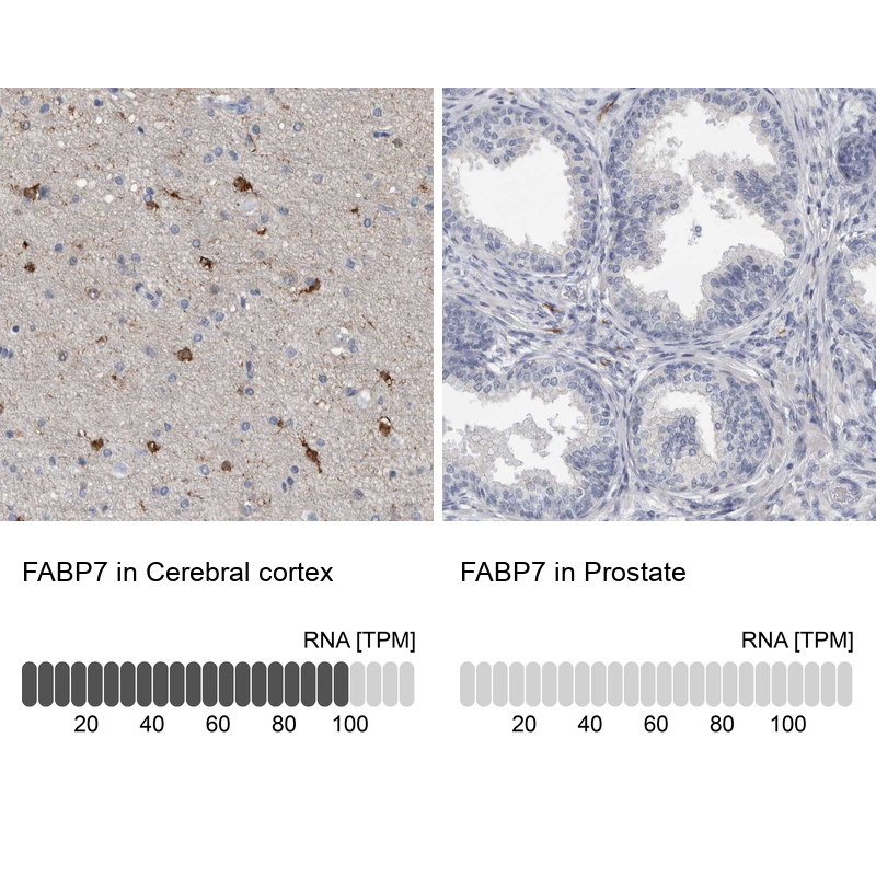
- Immunohistochemistry analysis in human cerebral cortex and prostate tissues using HPA028825 antibody. Corresponding FABP7 RNA-seq data are presented for the same tissues.
- Sample type
- Human
- Protocol
- Protocol