 Explore
 Validate
 Learn
Learn Western blot
Antibody data
- Antibody Data
- Antigen structure
- References [3]
- Comments [0]
- Validations
- Western blot [1]
Submit
Validation data
Reference
Comment
Report error
- Product number
- PAB10050 - Provider product page

- Provider
- Abnova Corporation
- Proper citation
- Abnova Corporation Cat#PAB10050, RRID:AB_1674923
- Product name
- GGA3 polyclonal antibody
- Antibody type
- Polyclonal
- Description
- Rabbit polyclonal antibody raised against synthetic peptide of GGA3.
- Storage
- Store at 4°C. For long term storage store at -20°C.Aliquot to avoid repeated freezing and thawing.
Submitted references Structural mechanism for ubiquitinated-cargo recognition by the Golgi-localized, gamma-ear-containing, ADP-ribosylation-factor-binding proteins.
The GAT domains of clathrin-associated GGA proteins have two ubiquitin binding motifs.
Structural basis for acidic-cluster-dileucine sorting-signal recognition by VHS domains.
Prag G, Lee S, Mattera R, Arighi CN, Beach BM, Bonifacino JS, Hurley JH
Proceedings of the National Academy of Sciences of the United States of America 2005 Feb 15;102(7):2334-9
Proceedings of the National Academy of Sciences of the United States of America 2005 Feb 15;102(7):2334-9
The GAT domains of clathrin-associated GGA proteins have two ubiquitin binding motifs.
Bilodeau PS, Winistorfer SC, Allaman MM, Surendhran K, Kearney WR, Robertson AD, Piper RC
The Journal of biological chemistry 2004 Dec 24;279(52):54808-16
The Journal of biological chemistry 2004 Dec 24;279(52):54808-16
Structural basis for acidic-cluster-dileucine sorting-signal recognition by VHS domains.
Misra S, Puertollano R, Kato Y, Bonifacino JS, Hurley JH
Nature 2002 Feb 21;415(6874):933-7
Nature 2002 Feb 21;415(6874):933-7
No comments: Submit comment
Supportive validation
- Submitted by
- Abnova Corporation (provider)
- Main image
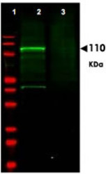
- Experimental details
- Western blot using GGA3 polyclonal antibody (Cat # PAB10050) shows detection of aband at ~110 KDa corresponding to GFP-GGA3 fusion protein present in a lysate of HEK293 cellsover- expressing the recombinant protein (Lane 2, arrowhead).Pre-incubation of antibody with immunizing peptide blocks specific staining (lane3).MW markers are shown in lane 1 (700 nm channel, red).Approximately 35 ug of lysate was separated on a 16% Tricine gel by SDS-PAGE and transferred onto nitrocellulose.After blocking the membrane was probed with the primary antibody diluted to 1:600.Reaction occurred overnight at 4°C followed by washes and reaction with a 1 : 10,000 dilution of IRDye™800 conjugated Gt-a-Rabbit IgG [H&L] for 45 min at room temperature (800 nm channel, green).IRDye™800 fluorescence image was captured using the Odyssey® Infrared Imaging System developed byLI-COR.IRDye is a trademark of LI-COR, Inc.
