Explore
 Validate
 Learn
Learn Western blot
 Immunocytochemistry
Antibody data
- Antibody Data
- Antigen structure
- References [7]
- Comments [0]
- Validations
- Immunocytochemistry [2]
- Immunohistochemistry [1]
- Flow cytometry [1]
- Other assay [1]
Submit
Validation data
Reference
Comment
Report error
- Product number
- MA5-11620 - Provider product page

- Provider
- Invitrogen Antibodies
- Product name
- Calponin 1 Monoclonal Antibody (CALP)
- Antibody type
- Monoclonal
- Antigen
- Other
- Description
- MA5-11620 targets Calponin-1 in IHC (P) applications and shows reactivity with Human and Rat samples. The MA5-11620 immunogen is crude human uterus extract.
- Reactivity
- Human, Mouse, Rat
- Host
- Mouse
- Isotype
- IgG
- Antibody clone number
- CALP
- Vial size
- 500 μL
- Concentration
- 0.2 mg/mL
- Storage
- 4°C
Submitted references Characterization of Burn Eschar Pericytes.
Chemerin-induced arterial contraction is G(i)- and calcium-dependent.
Pericardial synovial sarcoma, a potential for misdiagnosis: clinicopathologic and molecular cytogenetic analysis of three cases with literature review.
Metastatic inflammatory myofibroblastic tumor identified by EUS-FNA in mediastinal lymph nodes with ancillary FISH studies for ALK rearrangement.
TGF-beta1 drives partial myofibroblastic differentiation in chondromyxoid fibroma of bone.
Modulation of gene expression in precancerous rat esophagus by dietary zinc deficit and replenishment.
Identification of steroid sulfate transport processes in the human mammary gland.
Evdokiou A, Kanisicak O, Gierek S, Barry A, Ivey MJ, Zhang X, Bodnar RJ, Satish L
Journal of clinical medicine 2020 Feb 24;9(2)
Journal of clinical medicine 2020 Feb 24;9(2)
Chemerin-induced arterial contraction is G(i)- and calcium-dependent.
Ferland DJ, Darios ES, Neubig RR, Sjögren B, Truong N, Torres R, Dexheimer TS, Thompson JM, Watts SW
Vascular pharmacology 2017 Jan;88:30-41
Vascular pharmacology 2017 Jan;88:30-41
Pericardial synovial sarcoma, a potential for misdiagnosis: clinicopathologic and molecular cytogenetic analysis of three cases with literature review.
Cheng Y, Sheng W, Zhou X, Wang J
American journal of clinical pathology 2012 Jan;137(1):142-9
American journal of clinical pathology 2012 Jan;137(1):142-9
Metastatic inflammatory myofibroblastic tumor identified by EUS-FNA in mediastinal lymph nodes with ancillary FISH studies for ALK rearrangement.
Borak S, Siegal GP, Reddy V, Jhala N, Jhala D
Diagnostic cytopathology 2012 Aug;40 Suppl 2:E118-25
Diagnostic cytopathology 2012 Aug;40 Suppl 2:E118-25
TGF-beta1 drives partial myofibroblastic differentiation in chondromyxoid fibroma of bone.
Romeo S, Eyden B, Prins FA, Briaire-de Bruijn IH, Taminiau AH, Hogendoorn PC
The Journal of pathology 2006 Jan;208(1):26-34
The Journal of pathology 2006 Jan;208(1):26-34
Modulation of gene expression in precancerous rat esophagus by dietary zinc deficit and replenishment.
Liu CG, Zhang L, Jiang Y, Chatterjee D, Croce CM, Huebner K, Fong LY
Cancer research 2005 Sep 1;65(17):7790-9
Cancer research 2005 Sep 1;65(17):7790-9
Identification of steroid sulfate transport processes in the human mammary gland.
Pizzagalli F, Varga Z, Huber RD, Folkers G, Meier PJ, St-Pierre MV
The Journal of clinical endocrinology and metabolism 2003 Aug;88(8):3902-12
The Journal of clinical endocrinology and metabolism 2003 Aug;88(8):3902-12
No comments: Submit comment
Supportive validation
- Submitted by
- Invitrogen Antibodies (provider)
- Main image

- Experimental details
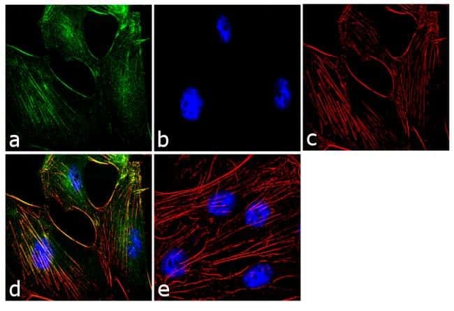
- Immunofluorescence analysis of Calponin-1 was performed using 70% confluent log phase MDA-MB-231 cells. The cells were fixed with 4% paraformaldehyde for 10 minutes, permeabilized with 0.1% Triton™ X-100 for 10 minutes, and blocked with 1% BSA for 1 hour at room temperature. The cells were labeled with Calponin-1 (CALP) Mouse Monoclonal Antibody (Product # MA5-11620) at 2 µg/mL in 0.1% BSA and incubated for 3 hours at room temperature and then labeled with Goat anti-Mouse IgG (H+L) Superclonal™ Secondary Antibody, Alexa Fluor® 488 conj µgate (Product # A28175) at a dilution of 1:2000 for 45 minutes at room temperature (Panel a: green). Nuclei (Panel b: blue) were stained with SlowFade® Gold Antifade Mountant with DAPI (Product # S36938). F-actin (Panel c: red) was stained with Alexa Fluor® 555 Rhodamine Phalloidin (Product # R415, 1:300). Panel d represents the merged image showing localization in the cytoskeleton. Panel e shows the no primary antibody control. The images were captured at 60X magnification.
- Submitted by
- Invitrogen Antibodies (provider)
- Main image
- Experimental details
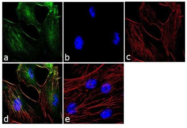
- Immunofluorescence analysis of Calponin-1 was performed using 70% confluent log phase MDA-MB-231 cells. The cells were fixed with 4% paraformaldehyde for 10 minutes, permeabilized with 0.1% Triton™ X-100 for 10 minutes, and blocked with 1% BSA for 1 hour at room temperature. The cells were labeled with Calponin-1 (CALP) Mouse Monoclonal Antibody (Product # MA5-11620) at 2 µg/mL in 0.1% BSA and incubated for 3 hours at room temperature and then labeled with Goat anti-Mouse IgG (H+L) Superclonal™ Secondary Antibody, Alexa Fluor® 488 conj µgate (Product # A28175) at a dilution of 1:2000 for 45 minutes at room temperature (Panel a: green). Nuclei (Panel b: blue) were stained with SlowFade® Gold Antifade Mountant with DAPI (Product # S36938). F-actin (Panel c: red) was stained with Alexa Fluor® 555 Rhodamine Phalloidin (Product # R415, 1:300). Panel d represents the merged image showing localization in the cytoskeleton. Panel e shows the no primary antibody control. The images were captured at 60X magnification.
Supportive validation
- Submitted by
- Invitrogen Antibodies (provider)
- Main image
- Experimental details
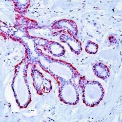
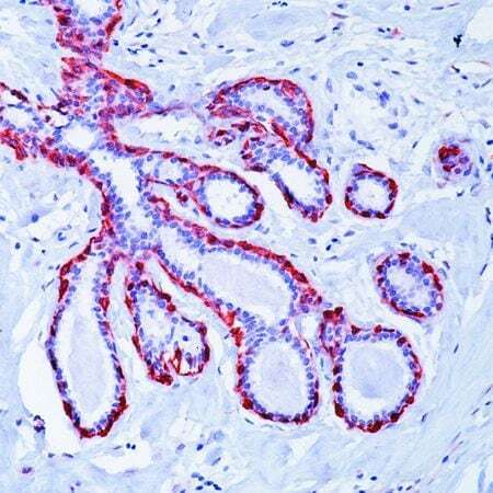
- Formalin-fixed, paraffin-embedded human breast stained with Calponin antibody using peroxidase-conjugate and AEC chromogen. Note cytoplasmic staining of myoepithelial cells in the lactiferous ducts.
Supportive validation
- Submitted by
- Invitrogen Antibodies (provider)
- Main image
- Experimental details
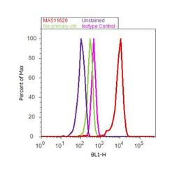
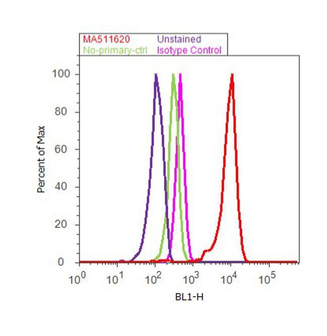
- Flow cytometry analysis of Calponin-1 was done on MCF7 cells. Cells were fixed with 70% ethanol for 10 minutes, permeabilized with 0.25% Triton™ X-100 for 20 minutes, and blocked with 5% BSA for 30 minutes at room temperature. Cells were labeled with Calponin-1 Mouse Monoclonal Antibody (Product # MA5-11620, red histogram) or with mouse isotype control (pink histogram) at 3-5 µg/million cells in 2.5% BSA. After incubation at room temperature for 2 hours, the cells were labeled with Alexa Fluor® 488 Rabbit Anti-Mouse Secondary Antibody (Product # A11059) at a dilution of 1:400 for 30 minutes at room temperature. The representative 10, 000 cells were acquired and analyzed for each sample using an Attune® Acoustic Focusing Cytometer. The purple histogram represents unstained control cells and the green histogram represents no-primary-antibody control.
Supportive validation
- Submitted by
- Invitrogen Antibodies (provider)
- Main image
- Experimental details
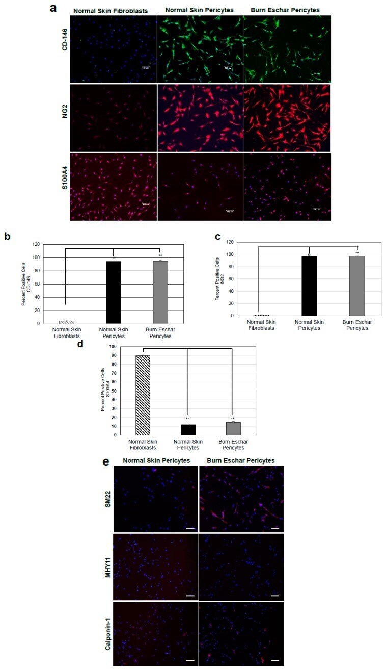
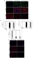
- Figure 2 Isolated cells are a heterogeneous population of pericytes. ( a ) Cultured pericytes from normal skin and burn eschar were analyzed for expression of the pericyte markers CD146 and NG2 along with the fibroblast marker S100A4. Representative images of the experiments performed on three different cultures derived from three different patients for normal skin pericytes, burn eschar pericytes and fibroblasts are shown. Images were captured using Eclipse 90i microscope and photographed with a DS-Qi1MC Digital Microscope. Scale bar: 100 muM. Quantitative analysis of ( b ) CD146, ( c ) NG2, and ( d ) S100A4 was performed. Quantitative analysis was performed in triplicate at 10x high powered fields using NIS-Elements AR3.1 software. Data represented as mean +- SEM of two independent studies. Statistical analysis was performed using Student's t test with p < 0.05 considered statistically significant. ** p < 0.01. The results show pericytes are greater than 90% positive for CD146 and NG2 and less than 15% positive for S100A4. The fibroblasts are greater than 90% positive for S100A4 and less than 10% positive for CD146 and NG2. ( e ) Shown is the representative images on the staining performed on normal skin pericytes and burn eschar pericytes derived from two different patients using SM22, myosin smooth muscle heavy chain (MHY)11, and Calponin-1 are shown. Scale bar: 30 uM.