Explore
 Validate
 Learn
Learn Western blot
 Immunohistochemistry
Antibody data
- Antibody Data
- Antigen structure
- References [2]
- Comments [0]
- Validations
- Immunohistochemistry [1]
Submit
Validation data
Reference
Comment
Report error
- Product number
- PA5-29725 - Provider product page

- Provider
- Invitrogen Antibodies
- Product name
- CTH Polyclonal Antibody
- Antibody type
- Polyclonal
- Antigen
- Recombinant full-length protein
- Description
- Recommended positive controls: 293T, A431, HeLa, HepG2, mouse liver, rat liver. Predicted reactivity: Mouse (84%), Rat (85%), Pig (86%), Bovine (91%). Store product as a concentrated solution. Centrifuge briefly prior to opening the vial.
- Reactivity
- Human, Mouse, Rat
- Host
- Rabbit
- Isotype
- IgG
- Vial size
- 100 μL
- Concentration
- 1.02 mg/mL
- Storage
- Store at 4°C short term. For long term storage, store at -20°C, avoiding freeze/thaw cycles.
Submitted references Hydrogen sulfide, oxygen, and calcium regulation in developing human airway smooth muscle.
Cystathionine metabolic enzymes play a role in the inflammation resolution of human keratinocytes in response to sub-cytotoxic formaldehyde exposure.
Bartman CM, Schiliro M, Helan M, Prakash YS, Linden D, Pabelick C
FASEB journal : official publication of the Federation of American Societies for Experimental Biology 2020 Sep;34(9):12991-13004
FASEB journal : official publication of the Federation of American Societies for Experimental Biology 2020 Sep;34(9):12991-13004
Cystathionine metabolic enzymes play a role in the inflammation resolution of human keratinocytes in response to sub-cytotoxic formaldehyde exposure.
Lee E, Kim HJ, Lee M, Jin SH, Hong SH, Ahn S, Kim SO, Shin DW, Lee ST, Noh M
Toxicology and applied pharmacology 2016 Nov 1;310:185-194
Toxicology and applied pharmacology 2016 Nov 1;310:185-194
No comments: Submit comment
Supportive validation
- Submitted by
- Invitrogen Antibodies (provider)
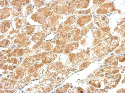
- Main image

- Experimental details
- CTH Polyclonal Antibody detects CTH protein at cytosol on human hepatoma by immunohistochemical analysis. Sample: Paraffin-embedded hepatoma tissue. CTH Polyclonal Antibody (Product # PA5-29725) dilution: 1:500. Antigen Retrieval: EDTA based buffer, pH 8.0, 15 min.