 Explore
 Validate
 Learn
LearnPA1-18296
antibody from Invitrogen Antibodies
Targeting: ATG16L1
APG16L, ATG16A, ATG16L, FLJ10035, WDR30
 Western blot
 Other assay
Antibody data
- Antibody Data
- Antigen structure
- References [1]
- Comments [0]
- Validations
- Other assay [1]
Submit
Validation data
Reference
Comment
Report error
- Product number
- PA1-18296 - Provider product page

- Provider
- Invitrogen Antibodies
- Product name
- ATG16L1 Polyclonal Antibody
- Antibody type
- Polyclonal
- Antigen
- Synthetic peptide
- Description
- Reconstitute in 100 µL of sterile water. Centrifuge to remove any insoluble material. After reconstitution keep aliquots at -20 °C for a higher stability, and at 4 °C with an appropriate antibacterial agent. Avoid repetitive freeze/thaw cycles.
- Reactivity
- Human, Rat
- Host
- Rabbit
- Isotype
- IgG
- Vial size
- 100 µL
- Concentration
- Conc. Not Determined
- Storage
- -20° C, Avoid Freeze/Thaw Cycles
Submitted references The Lipid Virulence Factors of Mycobacterium tuberculosis Exert Multilayered Control over Autophagy-Related Pathways in Infected Human Macrophages.
Bah A, Sanicas M, Nigou J, Guilhot C, Astarie-Dequeker C, Vergne I
Cells 2020 Mar 9;9(3)
Cells 2020 Mar 9;9(3)
No comments: Submit comment
Supportive validation
- Submitted by
- Invitrogen Antibodies (provider)
- Main image
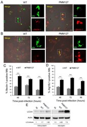
- Experimental details
- Figure 2 The Mtb mutant deficient in DIMs and SLs promoted autophagy activation. ( A - D ) Differentiated THP-1 cells were infected for 1 h with the indicated GFP-expressing Mtb strains; fixed at 48 h, 72 h, or 96 h postinfection (MOI between 5 and 10); permeabilized; incubated with antibodies against endogenous Beclin-1 ( A , C ) or Atg16L1 ( B , D ); and then stained with Alexa-568-labeled secondary antibody. Specimens were analyzed using confocal fluorescence microscopy. ( A , B ) Representative confocal images of macrophages infected with GFP-expressing Mtb (green channel) at 48 h postinfection, stained for Beclin-1 ( A ) or Atg16L1 ( B ) (red channel). The boxed areas in the left-hand panels are magnifications of the right-hand panels. Scale bars are 10 mum (left panels) or 2 mum (right panels). ( C , D ) A quantification of the percentage of Mtb compartments colocalized with Beclin-1 ( C ) or Atg16L1 ( D ) at 48 h, 72 h, and 96 h postinfection. Data are the mean +- s.e.m; 90 fields from three independent experiments; *** p < 0.0001 (unpaired t -test). ( E ) Differentiated THP-1 cells were infected for 1 h with the indicated Mtb strains and lysed 48 h postinfection. Cells were treated with 100 nM Bafilomycin A1 (with BafA1) or a DMSO (dimethylsulfoxide) (control (without BafA1) during the last 2 h postinfection. Lysates were analyzed using immunoblotting with anti-LC3 or anti-actin antibodies. The densitometric LC3-II/actin ratios are shown underneath the blot. The ratio
