PA5-20482
antibody from Invitrogen Antibodies
Targeting: ATG16L1
APG16L, ATG16A, ATG16L, FLJ10035, WDR30
Antibody data
- Antibody Data
- Antigen structure
- References [0]
- Comments [0]
- Validations
- Western blot [4]
- Immunocytochemistry [3]
Submit
Validation data
Reference
Comment
Report error
- Product number
- PA5-20482 - Provider product page
- Provider
- Invitrogen Antibodies
- Product name
- ATG16L1 Polyclonal Antibody
- Antibody type
- Polyclonal
- Antigen
- Synthetic peptide
- Description
- A suggested positive control is Hela cell lysate.
- Concentration
- 1 mg/mL
No comments: Submit comment
Supportive validation
- Submitted by
- Invitrogen Antibodies (provider)
- Main image
- Experimental details
- Western blot analysis of HeLa cell lysate using a ATG16 polyclonal antibody (Product # PA5-20482) at (A) 1 and (B) 2 µg/mL.
- Submitted by
- Invitrogen Antibodies (provider)
- Main image
- Experimental details
- Western Blot analysis of ATG16 in HeLa cell lysate with ATG16L1 Polyclonal Antibody (Product # PA5-20482) at (A) 1 and (B) 2 µg/mL.
- Submitted by
- Invitrogen Antibodies (provider)
- Main image
- Experimental details
- Knockdown of ATG16L1 was achieved by transfecting SH-SY5Y with ATG16L1 specific siRNAs (Silencer® select Product # S30069, S30070). Western Blot analysis (Fig. a) was performed using Whole cell extracts from the ATG16L1 knockdown cells (lane 3), non-targeting scrambled siRNA transfected cells (lane 2) and untransfected cells (lane 1). The blot was probed with ATG16L1 Polyclonal Antibody (Product # PA5-20482, 1 µg/mL ) and Goat anti-Rabbit IgG (H+L) Superclonal™ Recombinant Secondary Antibody, HRP (Product # A27036, 1:4000 dilution). Densitometric analysis of this Western Blot is shown in histogram (Fig. b). Decrease in signal upon siRNA mediated knock down confirms that antibody is specific to ATG16L1.
- Submitted by
- Invitrogen Antibodies (provider)
- Main image
- Experimental details
- Western Blot was performed using Anti-ATG16L1 Polyclonal Antibody (Product # PA5-20482) and a 64 kDa band corresponding to ATG16L1 was observed across cell lines tested. Whole cell extracts (30 µg lysate) of SH-SY5Y (Lane 1), PC-3 (Lane 2), MCF7 (Lane 3), HeLa (Lane 4) and HCT 116 (Lane 5) were electrophoresed using NuPAGE™ 4-12% Bis-Tris Protein Gel (Product # NP0322BOX). Resolved proteins were then transferred onto a nitrocellulose membrane (Product # IB23001) by iBlot® 2 Dry Blotting System (Product # IB21001). The blot was probed with the primary antibody (1 µg/mL) and detected by chemiluminescence with Goat anti-Rabbit IgG (H+L) Superclonal™ Recombinant Secondary Antibody, HRP (Product # A27036, 1:4000 dilution) using the iBright FL 1000 (Product # A32752). Chemiluminescent detection was performed using Novex® ECL Chemiluminescent Substrate Reagent Kit (Product # WP20005).
Supportive validation
- Submitted by
- Invitrogen Antibodies (provider)
- Main image
- Experimental details
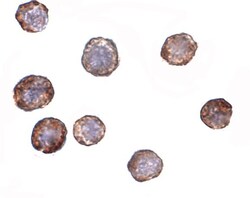
- Immunocytochemistry staining of HeLa cells using a ATG16 polyclonal antibody (Product # PA5-20482) at a 2 µg/mL dilution.
- Submitted by
- Invitrogen Antibodies (provider)
- Main image
- Experimental details
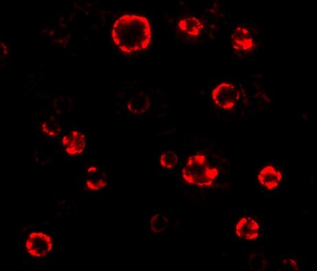
- Immunocytochemistry of ATG16 in HeLa cells with ATG16L1 Polyclonal Antibody (Product # PA5-20482) at 2 µg/mL.
- Submitted by
- Invitrogen Antibodies (provider)
- Main image
- Experimental details
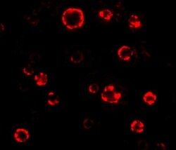
- Immunofluorescence of ATG16 in Hela cells with ATG16L1 Polyclonal Antibody (Product # PA5-20482) at 4.75 µg/mL.