 Explore
 Validate
 Learn
LearnPA5-48602
antibody from Invitrogen Antibodies
Targeting: RHBDF2
FLJ22341, iRhom2, RHBDL5, RHBDL6, TOC, TOCG
 Western blot
Antibody data
- Antibody Data
- Antigen structure
- References [1]
- Comments [0]
- Validations
- Western blot [3]
- Immunohistochemistry [4]
Submit
Validation data
Reference
Comment
Report error
- Product number
- PA5-48602 - Provider product page

- Provider
- Invitrogen Antibodies
- Product name
- RHBDF2 Polyclonal Antibody
- Antibody type
- Polyclonal
- Antigen
- Synthetic peptide
- Reactivity
- Human, Mouse
- Host
- Rabbit
- Isotype
- IgG
- Vial size
- 200 μL
- Concentration
- 0.5 mg/mL
- Storage
- Store at 4°C short term. For long term storage, store at -20°C, avoiding freeze/thaw cycles.
Submitted references Tyrosine phosphorylation directs TACE into extracellular vesicles via unconventional secretion.
Zhao Z, Kesti T, Uğurlu H, Baur AS, Fagerlund R, Saksela K
Traffic (Copenhagen, Denmark) 2019 Mar;20(3):202-212
Traffic (Copenhagen, Denmark) 2019 Mar;20(3):202-212
No comments: Submit comment
Supportive validation
- Submitted by
- Invitrogen Antibodies (provider)
- Main image

- Experimental details
- Western blot analysis of RHBDF2 in Mouse lung tissue lysate. Samples were incubated with RHBDF2 polyclonal antibody (Product # PA5-48602) using a dilution of 1:2,000 followed by Goat Anti-Rabbit IgG, (H+L), Peroxidase conjugated at a dilution of 1:10,000. Lysates/proteins: 20 µg per lane. Predicted band size: 97 kDa. Blocking/Dilution buffer: 5% NFDM/TBST.

- Submitted by
- Invitrogen Antibodies (provider)
- Main image
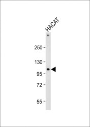
- Experimental details
- Western blot analysis of RHBDF2 in HACAT whole cell lysate. Samples were incubated with RHBDF2 polyclonal antibody (Product # PA5-48602) using a dilution of 1:2,000 followed by Goat Anti-Rabbit IgG, (H+L), Peroxidase conjugated at a dilution of 1:10,000. Lysates/proteins: 20 µg per lane. Predicted band size: 97 kDa. Blocking/Dilution buffer: 5% NFDM/TBST.

- Submitted by
- Invitrogen Antibodies (provider)
- Main image

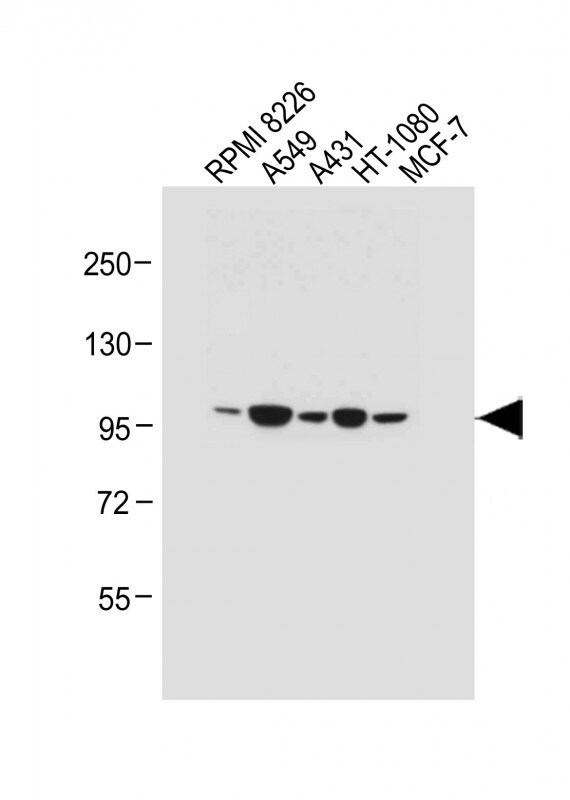
- Experimental details
- Western blot analysis of RHBDF2 in various lysates. Samples were incubated with RHBDF2 polyclonal antibody (Product # PA5-48602) using a dilution of 1:1,000 followed by Goat Anti-Rabbit IgG, (H+L), Peroxidase conjugated at a dilution of 1:10,000. Lysates/proteins: 20 µg per lane. Lane 1: RPMI 8226 whole cell lysate; Lane 2: A549 whole cell lysate; Lane 3: A431 whole cell lysate; Lane 4: HT-1080 whole cell lysate; Lane 5: MCF-7 whole cell lysate. Predicted band size: 97 kDa. Blocking/Dilution buffer: 5% NFDM/TBST.
Supportive validation
- Submitted by
- Invitrogen Antibodies (provider)
- Main image
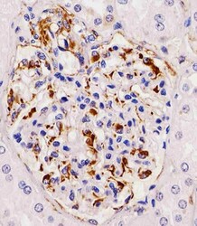
- Experimental details
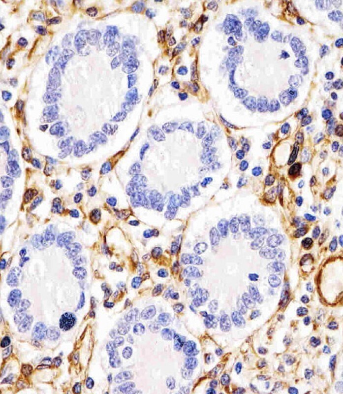
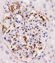
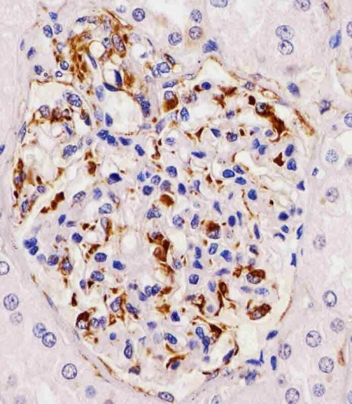
- Immunohistochemistry analysis of RHBDF2 in paraformaldehyde-fixed, paraffin-embedded human kidney tissue sections. Samples were incubated with RHBDF2 polyclonal antibody (Product # PA5-48602) using a dilution of 1:25 for 1 hours at 37°C followed by an undiluted biotinylated goat polyvalent antibody. Tissue was fixed with formaldehyde and blocked with 3% BSA for 0.5 hour at room temperature; antigen retrieval was by heat mediation with a citrate buffer (pH 6).
- Submitted by
- Invitrogen Antibodies (provider)
- Main image

- Experimental details
- Immunohistochemistry analysis of RHBDF2 in paraformaldehyde-fixed, paraffin-embedded human duodenum tissue sections. Samples were incubated with RHBDF2 polyclonal antibody (Product # PA5-48602) using a dilution of 1:25 for 1 hours at 37°C followed by an undiluted biotinylated goat polyvalent antibody. Tissue was fixed with formaldehyde and blocked with 3% BSA for 0.5 hour at room temperature; antigen retrieval was by heat mediation with a citrate buffer (pH 6).
- Submitted by
- Invitrogen Antibodies (provider)
- Main image
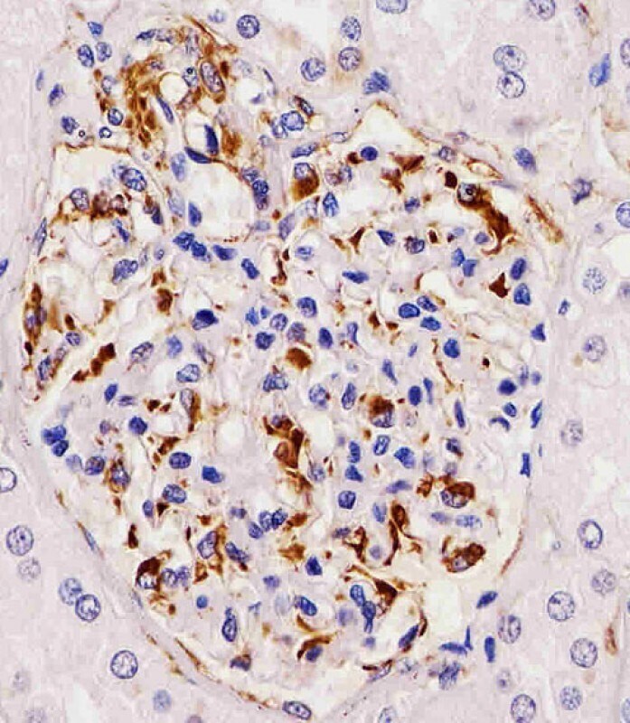
- Experimental details
- Immunohistochemistry analysis of RHBDF2 in paraformaldehyde-fixed, paraffin-embedded human kidney tissue sections. Samples were incubated with RHBDF2 polyclonal antibody (Product # PA5-48602) using a dilution of 1:25 for 1 hours at 37°C followed by an undiluted biotinylated goat polyvalent antibody. Tissue was fixed with formaldehyde and blocked with 3% BSA for 0.5 hour at room temperature; antigen retrieval was by heat mediation with a citrate buffer (pH 6).
- Submitted by
- Invitrogen Antibodies (provider)
- Main image

- Experimental details
- Immunohistochemistry analysis of RHBDF2 in paraformaldehyde-fixed, paraffin-embedded human duodenum tissue sections. Samples were incubated with RHBDF2 polyclonal antibody (Product # PA5-48602) using a dilution of 1:25 for 1 hours at 37°C followed by an undiluted biotinylated goat polyvalent antibody. Tissue was fixed with formaldehyde and blocked with 3% BSA for 0.5 hour at room temperature; antigen retrieval was by heat mediation with a citrate buffer (pH 6).
