 Explore
 Validate
 Learn
LearnPA5-22731
antibody from Invitrogen Antibodies
Targeting: MAP1LC3A
ATG8E, LC3, LC3A, MAP1ALC3, MAP1BLC3
 Western blot
 ELISA
ELISAAntibody data
- Antibody Data
- Antigen structure
- References [0]
- Comments [0]
- Validations
- Western blot [1]
- Other assay [1]
Submit
Validation data
Reference
Comment
Report error
- Product number
- PA5-22731 - Provider product page

- Provider
- Invitrogen Antibodies
- Product name
- LC3A/LC3B Polyclonal Antibody, DyLight™ 488
- Antibody type
- Polyclonal
- Antigen
- Synthetic peptide
- Description
- The target sequence has 100% sequence homology with xenopus, bovine and zebrafish.
- Reactivity
- Human, Mouse, Rat, Canine, Zebrafish
- Host
- Rabbit
- Conjugate
- Green dye
- Isotype
- IgG
- Vial size
- 100 µL
- Concentration
- 0.78 mg/mL
- Storage
- 4° C, store in dark
No comments: Submit comment
Supportive validation
- Submitted by
- Invitrogen Antibodies (provider)
- Main image

- Experimental details
- Western blot was performed using Anti-LC3A/LC3B Rabbit Polyclonal Antibody (Product # PA5-22731) and 14-16 kDa bands corresponding to LC3B were observed across cell lines tested and increased upon Chloroquine treatment. Whole cell extracts (30 µg lysate) of NIH3T3 (Lane 1), NIH3T3 treated with Chloroquine (50uM for 12 Hours) (Lane 2), HCT 116 (Lane 3) and HCT 116 treated with Chloroquine (50uM for 12 Hours) (Lane 4) were electrophoresed using Novex® NuPAGE® 12 % Bis-Tris gel (Product # NP0342BOX). Resolved proteins were then transferred onto a nitrocellulose membrane (Product #) by iBlot® 2 Dry Blotting System (Product # IB21001). The bot was probed with the primary antibody (1µg/mL) and detected by chemiluminescence with Goat anti-Rabbit IgG (H+L) Superclonal™ Recombinant Secondary Antibody, HRP (Product # A27036, 1:4000 dilution) using the iBright FL 1000 (Product # A32752). Chemiluminescent detection was performed using Novex® ECL Chemiluminescent Substrate Reagent Kit (Product # WP20005).
- Conjugate
- Green dye

Supportive validation
- Submitted by
- Invitrogen Antibodies (provider)
- Main image
- Experimental details
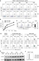
- Figure 5 AMPK deficiency promotes CD8 T cell death during activation A, cells from LNs of WT and AMPK KO mice were stimulated with PMA (10ng/ml) plus indicated concentrations of ionomycin for 6 hours. Flow cytometric staining for 7-AAD and Annexin V in CD8 + T cells. B, cells from LNs of WT and KO mice were stimulated with PMA (10ng/ml)/ionomycin (500ng/ml) for indicated time periods. CD8 + T cell death was analyzed by flow cytometric staining of 7-AAD (*, p
- Conjugate
- Green dye
