 Explore
 Validate
 Learn
Learn Western blot
Antibody data
- Antibody Data
- Antigen structure
- References [4]
- Comments [0]
- Validations
- Western blot [5]
Submit
Validation data
Reference
Comment
Report error
- Product number
- AP3301a - Provider product page

- Provider
- Abgent
- Proper citation
- Abgent Cat#AP3301a, RRID:AB_2137560
- Product name
- Phospho-LC3C(S12) Antibody
- Antibody type
- Polyclonal
- Antigen
- Synthetic peptide
- Description
- Peptide Affinity Purified Rabbit Polyclonal Antibody (Pab)
- Reactivity
- Human
- Host
- Rabbit
- Isotype
- IgG
- Vial size
- 400 µl
- Concentration
- 0.5 mg/ml
- Storage
- Maintain refrigerated at 2-8°C for up to 6 months. For long term storage store at -20°C in small aliquots to prevent freeze-thaw cycles.
Submitted references Identification of a small molecule targeting annexin A7.
MAPK15/ERK8 stimulates autophagy by interacting with LC3 and GABARAP proteins.
Rab5 and class III phosphoinositide 3-kinase Vps34 are involved in hepatitis C virus NS4B-induced autophagy.
Regulation of the autophagy protein LC3 by phosphorylation.
Li H, Liu N, Wang S, Wang L, Zhao J, Su L, Zhang Y, Zhang S, Xu Z, Zhao B, Miao J
Biochimica et biophysica acta 2013 Sep;1833(9):2092-9
Biochimica et biophysica acta 2013 Sep;1833(9):2092-9
MAPK15/ERK8 stimulates autophagy by interacting with LC3 and GABARAP proteins.
Colecchia D, Strambi A, Sanzone S, Iavarone C, Rossi M, Dall'Armi C, Piccioni F, Verrotti di Pianella A, Chiariello M
Autophagy 2012 Dec;8(12):1724-40
Autophagy 2012 Dec;8(12):1724-40
Rab5 and class III phosphoinositide 3-kinase Vps34 are involved in hepatitis C virus NS4B-induced autophagy.
Su WC, Chao TC, Huang YL, Weng SC, Jeng KS, Lai MM
Journal of virology 2011 Oct;85(20):10561-71
Journal of virology 2011 Oct;85(20):10561-71
Regulation of the autophagy protein LC3 by phosphorylation.
Cherra SJ 3rd, Kulich SM, Uechi G, Balasubramani M, Mountzouris J, Day BW, Chu CT
The Journal of cell biology 2010 Aug 23;190(4):533-9
The Journal of cell biology 2010 Aug 23;190(4):533-9
No comments: Submit comment
Supportive validation
- Submitted by
- Abgent (provider)
- Main image
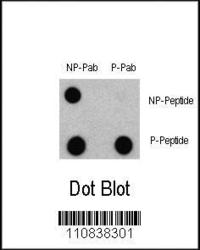
- Experimental details
- Dot blot analysis of Phospho-LC3 (APG8a) - S12 Antibody (Cat. #AP3301a) and Nonphospho-LC3 (APG8a) Antibody on nitrocellulose membrane. 50ng of Phospho-peptide or Non Phospho-peptide per dot were adsorbed. Antibody working concentrations are 0.5ug per ml.
- Primary Ab dilution
- 1:500

- Submitted by
- Abgent (provider)
- Main image

- Experimental details
- "Immunoblots of phosphorylated LC3 (phospho-LC3) in CHO cell culture. LC3 and LC3 S12A mutant vectors were transfected into CHO cells. The cell lysates were separated with SDS-PAGE and blotted with anti-phospho-LC3 S12 antibody. LC3 = microtubule-associated protein light chain-3; S12A = replacement of the amino acid position 12 serine of LC3 with alanine. WT = wildtype LC3-transfected cell lysates; S12A = LC3 S12A mutant-transfected cell lysates; Empty vector = vector with no LC3 gene. Molecular size: LC3-I = 16kDa, and LC3-II = 14 kDa"
- Primary Ab dilution
- 1:1000

- Submitted by
- Abgent (provider)
- Main image

- Experimental details
- Immunoblots of SH-SY5Y cells treated with rapamycin for 1 h was probed with AP3301a. The data shows that treatment with rapamycin showed no significant change in level of LC3.
- Primary Ab dilution
- 1:1000

- Submitted by
- Abgent (provider)
- Main image

- Experimental details
- Something like SH-SY5Y cells expressing GFP-LC3-WT or-S12D treated with rapamycin or vehicle for 1h.
- Primary Ab dilution
- 1:1000

- Submitted by
- Abgent (provider)
- Main image

- Experimental details
- Immunoblots of SH-SY5Y cells treated with MPP+ for 24h was probed with AP3301a. The data shows that treatment with MPP+ showed no significant change in level of LC3.
- Primary Ab dilution
- 1:1000
