Explore
 Validate
 Learn
LearnA01543-1
antibody from Boster Biological Technology
Targeting: MAP1LC3A
ATG8E, LC3, LC3A, MAP1ALC3, MAP1BLC3
 Western blot
 Immunohistochemistry
Antibody data
- Antibody Data
- Antigen structure
- References [3]
- Comments [0]
- Validations
- Immunohistochemistry [1]
Submit
Validation data
Reference
Comment
Report error
- Product number
- A01543-1 - Provider product page

- Provider
- Boster Biological Technology
- Product name
- Anti-MAP1LC3A Antibody Picoband™
- Antibody type
- Polyclonal
- Description
- Rabbit IgG polyclonal antibody for MAP1LC3A detection. Tested with WB, IHC-P, Direct ELISA in Human;Mouse;Rat.
- Reactivity
- Human, Mouse, Rat
- Host
- Rabbit
- Vial size
- 100μg/vial
- Concentration
- 0.5-1mg/ml, actual concentration vary by lot. Use suggested dilution ratio to decide dilution procedure.
- Storage
- At -20°C for one year. After reconstitution, at 4°C for one month. It can also be aliquoted and stored frozen at -20°C for a longer time. Avoid repeated freezing and thawing.
- Handling
- Add 0.2ml of distilled water will yield a concentration of 500ug/ml.
Submitted references Circ_0002331 Interacts with ELAVL1 to Improve ox-LDL-Induced Vascular Endothelial Cell Dysfunction via Regulating CCND2 mRNA Stability.
Polyphyllin I Promotes Autophagic Cell Death and Apoptosis of Colon Cancer Cells via the ROS-Inhibited AKT/mTOR Pathway.
Association of astragaloside IV-inhibited autophagy and mineralization in vascular smooth muscle cells with lncRNA H19 and DUSP5-mediated ERK signaling.
Chen F, Yu X
Cardiovascular toxicology 2024 Jul;24(7):625-636
Cardiovascular toxicology 2024 Jul;24(7):625-636
Polyphyllin I Promotes Autophagic Cell Death and Apoptosis of Colon Cancer Cells via the ROS-Inhibited AKT/mTOR Pathway.
Luo Q, Jia L, Huang C, Qi Q, Jahangir A, Xia Y, Liu W, Shi R, Tang L, Chen Z
International journal of molecular sciences 2022 Aug 19;23(16)
International journal of molecular sciences 2022 Aug 19;23(16)
Association of astragaloside IV-inhibited autophagy and mineralization in vascular smooth muscle cells with lncRNA H19 and DUSP5-mediated ERK signaling.
Song Z, Wei D, Chen Y, Chen L, Bian Y, Shen Y, Chen J, Pan Y
Toxicology and applied pharmacology 2019 Feb 1;364:45-54
Toxicology and applied pharmacology 2019 Feb 1;364:45-54
No comments: Submit comment
Supportive validation
- Submitted by
- Boster Biological Technology (provider)
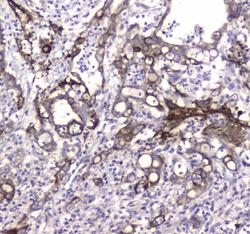
- Main image
- Experimental details
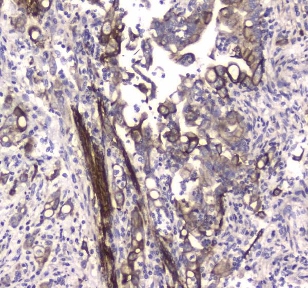
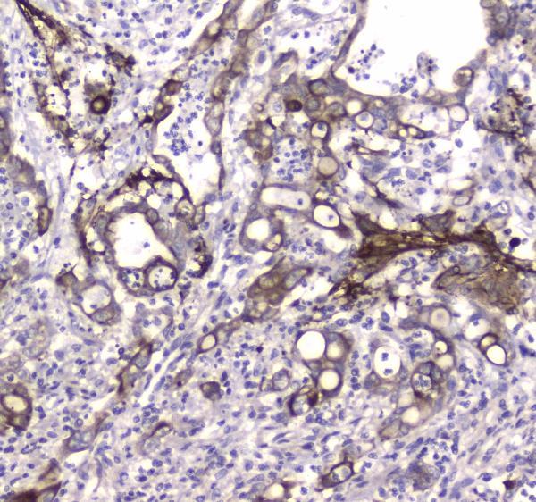
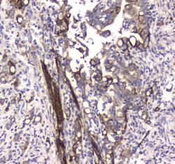
- IHC analysis of MAP1LC3A using anti-MAP1LC3A antibody (A01543-1). MAP1LC3A was detected in paraffin-embedded section of human colon cancer tissue. Heat mediated antigen retrieval was performed in citrate buffer (pH6, epitope retrieval solution) for 20 mins. The tissue section was blocked with 10% goat serum. The tissue section was then incubated with 1μg/ml rabbit anti-MAP1LC3A Antibody (A01543-1) overnight at 4°C. Biotinylated goat anti-rabbit IgG was used as secondary antibody and incubated for 30 minutes at 37°C. The tissue section was developed using Strepavidin-Biotin-Complex (SABC)(Catalog # SA1022) with DAB as the chromogen.
- Additional image