Explore
 Validate
 Learn
Learn Western blot
Antibody data
- Antibody Data
- Antigen structure
- References [2]
- Comments [0]
- Validations
- Western blot [2]
- Immunohistochemistry [1]
Submit
Validation data
Reference
Comment
Report error
- Product number
- AP1813b - Provider product page

- Provider
- Abgent
- Proper citation
- Abgent Cat#AP1813b, RRID:AB_2062173
- Product name
- ATG7 Antibody (Center)
- Antibody type
- Polyclonal
- Antigen
- Synthetic peptide
- Description
- Purified Rabbit Polyclonal Antibody (Pab)
- Reactivity
- Human, Mouse
- Host
- Rabbit
- Isotype
- IgG
- Vial size
- 400 µl
- Concentration
- 2 mg/ml
- Storage
- Maintain refrigerated at 2-8°C for up to 6 months. For long term storage store at -20°C in small aliquots to prevent freeze-thaw cycles.
Submitted references Autophagy deficiency by hepatic FIP200 deletion uncouples steatosis from liver injury in NAFLD.
Temporal orchestration of circadian autophagy rhythm by C/EBPβ.
Ma D, Molusky MM, Song J, Hu CR, Fang F, Rui C, Mathew AV, Pennathur S, Liu F, Cheng JX, Guan JL, Lin JD
Molecular endocrinology (Baltimore, Md.) 2013 Oct;27(10):1643-54
Molecular endocrinology (Baltimore, Md.) 2013 Oct;27(10):1643-54
Temporal orchestration of circadian autophagy rhythm by C/EBPβ.
Ma D, Panda S, Lin JD
The EMBO journal 2011 Sep 6;30(22):4642-51
The EMBO journal 2011 Sep 6;30(22):4642-51
No comments: Submit comment
Supportive validation
- Submitted by
- Abgent (provider)
- Main image

- Experimental details
- Western blot analysis of anti-ATG7 Antibody (Center) Pab (Cat. #AP1813b) in 293 cell line lysates transiently transfected with the ATG7 gene (2ug/lane). hAPG7L-P299(arrow) was detected using the purified Pab.
- Primary Ab dilution
- 1:1000

- Submitted by
- Abgent (provider)
- Main image

- Experimental details
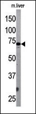
- The ATG7 Antibody (Center) Pab (Cat. #AP1813b) is used in Western blot to detect APG7L in mouse liver tissue lysate. APG7L (arrow) was detected using the purified Pab.
- Primary Ab dilution
- 1:1000
Supportive validation
- Submitted by
- Abgent (provider)
- Main image

- Experimental details
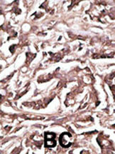
- "Formalin-fixed and paraffin-embedded human cancer tissue reacted with the primary antibody, which was peroxidase-conjugated to the secondary antibody, followed by DAB staining. This data demonstrates the use of this antibody for immunohistochemistry; clinical relevance has not been evaluated. BC = breast carcinoma; HC = hepatocarcinoma."
- Primary Ab dilution
- 1:50~100