 Explore
 Validate
 Learn
Learn Western blot
Antibody data
- Antibody Data
- Antigen structure
- References [0]
- Comments [0]
- Validations
- Western blot [3]
- Immunocytochemistry [1]
- Immunohistochemistry [1]
Submit
Validation data
Reference
Comment
Report error
- Product number
- TA324635 - Provider product page

- Provider
- OriGene
- Product name
- Rabbit polyclonal ATG7 Antibody
- Antibody type
- Polyclonal
- Description
- Rabbit polyclonal ATG7 Antibody
- Host
- Rabbit
- Conjugate
- Unconjugated
- Epitope
- ATG7
- Isotype
- IgG
- Antibody clone number
- NULL
- Vial size
- 400 µl
- Concentration
- 0.5 mg/ml
No comments: Submit comment
Supportive validation
- Submitted by
- OriGene (provider)
- Main image
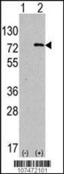
- Experimental details
- Western blot analysis of anti-hAPG7L-R509 Pab (Cat. #TA324635) in 293 cell line lysates transiently transfected with the ATG7 gene (2ug/lane). hAPG7L-R509(arrow) was detected using the purified Pab.
- Validation comment
- WB
- Submitted by
- OriGene (provider)
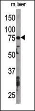
- Main image

- Experimental details
- The anti-APG7L Pab (Cat. #TA324635) is used in Western blot to detect APG7L in mouse liver tissue lysate. APG7L(arrow) was detected using the purified Pab.
- Validation comment
- WB
- Submitted by
- OriGene (provider)
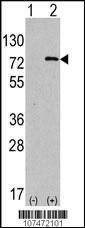
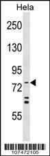
- Main image

- Experimental details
- APG7L Antibody (R509) (Cat. #TA324635) western blot analysis in Hela cell line lysates (35ug/lane).This demonstrates the APG7L antibody detected the APG7L protein (arrow).
- Validation comment
- WB
Supportive validation
- Submitted by
- OriGene (provider)
- Main image

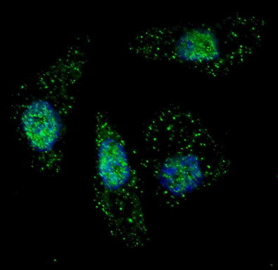
- Experimental details
- IF image of U251 cells stained with ATG7 antibody. U251 cells were treated with Chloroquine , then incubated with TA324635 ATG7 primary antibody (1:200, 2 h at RT). For secondary antibody, Alexa Fluor? 488 conjugated donkey anti-rabbit antibody (green) was used (1:1000, 1h). Nuclei were counterstained with Hoechst 33342 (blue) . ATG7 immunoreactivity is localized to autophagic vacuoles in the cytoplasm of U251 cells.
- Validation comment
- IF
Supportive validation
- Submitted by
- OriGene (provider)
- Main image
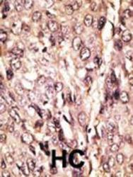
- Experimental details
- Formalin-fixed and paraffin-embedded human cancer tissue reacted with the primary antibody, which was peroxidase-conjugated to the secondary antibody, followed by AEC staining. This data demonstrates the use of this antibody for immunohistochemistry; clinical relevance has not been evaluated. BC = breast carcinoma; HC = hepatocarcinoma.
- Validation comment
- IHC
