Explore
 Validate
 Learn
Learn Western blot
 Immunoprecipitation
Antibody data
- Antibody Data
- Antigen structure
- References [5]
- Comments [0]
- Validations
- Western blot [3]
- Other assay [5]
Submit
Validation data
Reference
Comment
Report error
- Product number
- PA5-17216 - Provider product page

- Provider
- Invitrogen Antibodies
- Product name
- ATG7 Polyclonal Antibody
- Antibody type
- Polyclonal
- Antigen
- Synthetic peptide
- Description
- It is not recommended to aliquot this antibody.
- Reactivity
- Human, Mouse, Rat
- Host
- Rabbit
- Isotype
- IgG
- Vial size
- 100 µL
- Concentration
- 8 µg/mL
- Storage
- -20°C
Submitted references Knockdown of VDAC1 alleviates the cognitive dysfunction secondary to sepsis-associated encephalopathy.
Vasorin/ATIA Promotes Cigarette Smoke-Induced Transformation of Human Bronchial Epithelial Cells by Suppressing Autophagy-Mediated Apoptosis.
Synergistic anticancer effect of cisplatin and Chal-24 combination through IAP and c-FLIPL degradation, Ripoptosome formation and autophagy-mediated apoptosis.
A JNK-mediated autophagy pathway that triggers c-IAP degradation and necroptosis for anticancer chemotherapy.
Attenuation of TNFSF10/TRAIL-induced apoptosis by an autophagic survival pathway involving TRAF2- and RIPK1/RIP1-mediated MAPK8/JNK activation.
Cai M, Du B, Si Y, Miao J, Ge J, Zhang J, Song J, Bao H
American journal of translational research 2021;13(7):7538-7555
American journal of translational research 2021;13(7):7538-7555
Vasorin/ATIA Promotes Cigarette Smoke-Induced Transformation of Human Bronchial Epithelial Cells by Suppressing Autophagy-Mediated Apoptosis.
Chen W, Wang Q, Xu X, Saxton B, Tessema M, Leng S, Choksi S, Belinsky SA, Liu ZG, Lin Y
Translational oncology 2020 Jan;13(1):32-41
Translational oncology 2020 Jan;13(1):32-41
Synergistic anticancer effect of cisplatin and Chal-24 combination through IAP and c-FLIPL degradation, Ripoptosome formation and autophagy-mediated apoptosis.
Shi S, Wang Q, Xu J, Jang JH, Padilla MT, Nyunoya T, Xing C, Zhang L, Lin Y
Oncotarget 2015 Jan 30;6(3):1640-51
Oncotarget 2015 Jan 30;6(3):1640-51
A JNK-mediated autophagy pathway that triggers c-IAP degradation and necroptosis for anticancer chemotherapy.
He W, Wang Q, Srinivasan B, Xu J, Padilla MT, Li Z, Wang X, Liu Y, Gou X, Shen HM, Xing C, Lin Y
Oncogene 2014 Jun 5;33(23):3004-13
Oncogene 2014 Jun 5;33(23):3004-13
Attenuation of TNFSF10/TRAIL-induced apoptosis by an autophagic survival pathway involving TRAF2- and RIPK1/RIP1-mediated MAPK8/JNK activation.
He W, Wang Q, Xu J, Xu X, Padilla MT, Ren G, Gou X, Lin Y
Autophagy 2012 Dec;8(12):1811-21
Autophagy 2012 Dec;8(12):1811-21
No comments: Submit comment
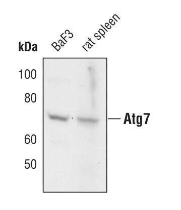
Supportive validation
- Submitted by
- Invitrogen Antibodies (provider)
- Main image
- Experimental details
- Western blot analysis of Atg7 was performed by loading 20 µg of HEK293T whole cell lysate per well onto a SDS-PAGE gel. Proteins were transferred to a PVDF membrane and blocked with 5% non-fat dry milk in TBST for 1 hour at room temperature. The membrane was probed with an Atg7 polyclonal antibody (Product # PA5-17216) at a dilution of 1:1000 overnight at 4°C, washed in TBST, and probed with an HRP-conjugated goat anti-rabbit IgG secondary antibody at a dilution of 1:40,000 for 1 hour at room temperature. Chemiluminescent detection was performed using ECL substrate. Data courtesy of the Innovators Program.

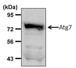
- Submitted by
- Invitrogen Antibodies (provider)
- Main image

- Experimental details
- Western blot analysis of Atg7 in extracts from BaF3 cells and rat spleen using Atg7 polyclonal antibody (Product # PA5-17216).
- Submitted by
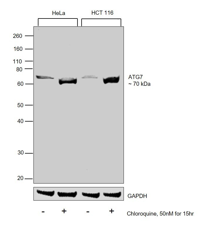
- Invitrogen Antibodies (provider)
- Main image

- Experimental details
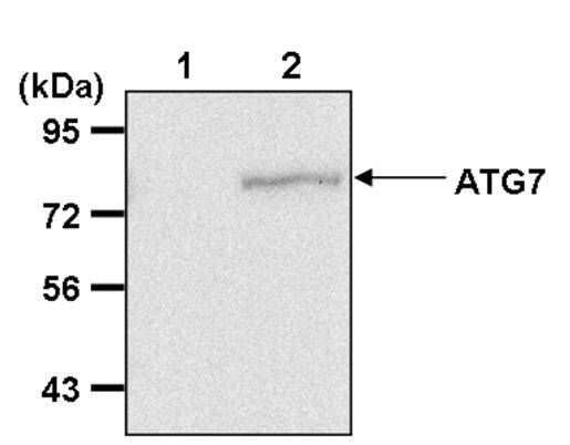
- Western blot was performed using Anti-ATG7 Polyclonal Antibody (Product # PA5-17216) and a 70 kDa band corresponding to ATG7 was observed across cell lines tested and increased upon Chloroquine treatment. Whole cell extracts (30 µg lysate) of HeLa (Lane 1), HeLa treated with Chloroquine (50nM for 15 hr) (Lane 2), HCT 116 (Lane 3) and HCT 116 treated with Chloroquine (50nM for 15 hr) (Lane 4) were electrophoresed using Novex® NuPAGE® 12 % Bis-Tris gel (Product # NP0342BOX). Resolved proteins were then transferred onto a nitrocellulose membrane (Product # IB23001) by iBlot® 2 Dry Blotting System (Product # IB21001). The blot was probed with the primary antibody (1:1000 dilution) and detected by chemiluminescence with Goat anti-Rabbit IgG (H+L), Superclonal™ Recombinant Secondary Antibody, HRP (Product # A27036, 1:4000 dilution) using the iBright FL 1000 (Product # A32752). Chemiluminescent detection was performed using Novex® ECL Chemiluminescent Substrate Reagent Kit (Product # WP20005).
Supportive validation
- Submitted by
- Invitrogen Antibodies (provider)
- Main image

- Experimental details
- Immunoprecipitation of Atg7 was performed on HEK293T cells. Antigen-antibody complexes were formed by incubating 500 µg of HEK293T whole cell lysate (in 300 µL volume) with 3 µL of an Atg7 polyclonal antibody (Product # PA5-17216) (Lane 2) or a rabbit IgG isotype control (Lane 1) overnight at 4°C. The immune complexes were captured on 30 µL of protein G-sepharose, washed extensively, and eluted with 6X Laemmli buffer. Samples were resolved on an 8% SDS-PAGE, transferred to a PVDF membrane, and blocked with 5% milk in TBST for 1 hour at room temperature. The membrane was probed with an Atg7 polyclonal antibody (Product # PA5-17216) at a dilution of 1:1000 overnight at 4°C, washed in TBST, and probed with an HRP-conjugated goat anti-rabbit light chain secondary antibody at a dilution of 1:40,000 for 1 hour at room temperature. Chemiluminescent detection was performed using ECL substrate. Data courtesy of the Innovators Program.
- Submitted by
- Invitrogen Antibodies (provider)
- Main image
- Experimental details
- NULL

- Submitted by
- Invitrogen Antibodies (provider)
- Main image
- Experimental details
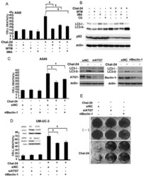
- Fig 4 Chal-24 induces cell death depending on Autophagy (A) A549 cells were pretreated with 3MA (10 mM), CQ (20 muM), or Wortmannin (WTM, 1 muM) for 30 min and then treated with Chal-24 (8 muM) for an additional 30 h. Cytotoxicity was detected by LDH release assay. Data shown are the mean +- SD. * p

- Submitted by
- Invitrogen Antibodies (provider)
- Main image

- Experimental details
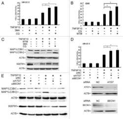
- Figure 2 Autophagy is required for cell death induced by Chal-24 and cisplatin combination (A) A549 cells were treated with cisplatin (10 muM), Chal-24 (1 muM) alone or in combination for the indicated times. The indicated proteins were detected by Western blot. beta-tubulin was used as an input control. (B) the cells were preteated with chloroquine (CQ, 20 muM) for 30 min, and then treated with cisplatin and Chal-24 (24 h or 4 h for upper panel for p62 detection and lower panel for LC3 detection, respectively). The indicated proteins were detected by Western blot. GAPDH was detected as an input control. (C) A549 cells were pretreated with autophagy inhibitors (CQ, 20 muM; WTM, 1 muM; 3MA, 10 muM) for 30 min, followed by cisplatin (10 muM) and Chal-24 (1 muM) co-treatment for an additional 48 h, cell death was measured as described in Figure 1 . (D) the cells were transfected with the indicated siRNA for 24 h, then the cells were treated with cisplatin (10 muM) and Chal-24 (1 muM) for 48 h, cell death was measured by LDH assay. * p < 0.05, ** p < 0.01. Insert, knockdown of ATG7 expression was confirmed by Western blot.
- Submitted by
- Invitrogen Antibodies (provider)
- Main image
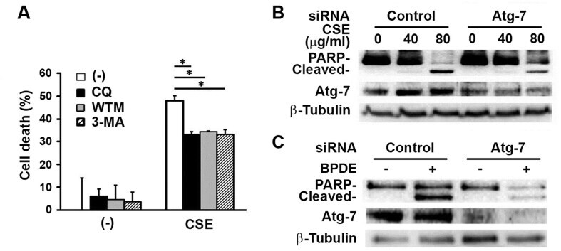
- Experimental details
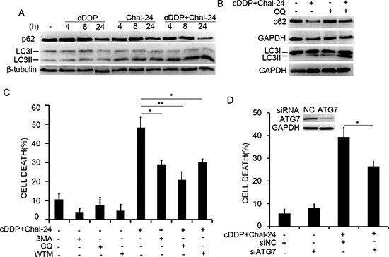
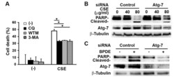
- Figure 5 Autophagy contributes to CSE-induced cell death . (A) BEAS-2B cells treated with autophagy inhibitors CQ (20 muM), WTM (1 muM) or 3-MA (10 mM) for 30 min followed by incubation of CSE (20 mug/mL) for 48 hours. Cell death was determined in triplicate samples by LDH release assay. * P < 0.05. (B, C) BEAS-2B cells were transfected with control or Atg-7 siRNA (10 nM) for 24 hours, then treated with CSE (20 mug/mL, B) or BPDE (0.4 muM) for 4 hours (C). PARP cleavage and Atg-7 levels were examined by western blot with beta-tubulin as a loading control. Figure 5