 Explore
 Validate
 Learn
Learn Western blot
 Immunocytochemistry
Antibody data
- Antibody Data
- Antigen structure
- References [4]
- Comments [0]
- Validations
- Immunocytochemistry [1]
- Immunohistochemistry [1]
Submit
Validation data
Reference
Comment
Report error
- Product number
- HPA012783 - Provider product page

- Provider
- Atlas Antibodies
- Proper citation
- Atlas Antibodies Cat#HPA012783, RRID:AB_1847843
- Product name
- Anti-DPEP1
- Antibody type
- Polyclonal
- Description
- Polyclonal Antibody against Human DPEP1, Gene description: dipeptidase 1 (renal), Validated applications: ICC, WB, IHC, Uniprot ID: P16444, Storage: Store at +4°C for short term storage. Long time storage is recommended at -20°C.
- Reactivity
- Human
- Host
- Rabbit
- Conjugate
- Unconjugated
- Isotype
- IgG
- Vial size
- 100 µl
- Concentration
- 0.1 mg/ml
- Storage
- Store at +4°C for short term storage. Long time storage is recommended at -20°C.
- Handling
- The antibody solution should be gently mixed before use.
Submitted references A human stomach cell type transcriptome atlas
Supermeres are functional extracellular nanoparticles replete with disease biomarkers and therapeutic targets
Clinicopathological examination of dipeptidase�1 expression in colorectal cancer
Distinguishing primary from secondary mucinous ovarian tumors: an algorithm using the novel marker DPEP1
Öling S, Struck E, Noreen-Thorsen M, Zwahlen M, von Feilitzen K, Odeberg J, Pontén F, Lindskog C, Uhlén M, Dusart P, Butler L
BMC Biology 2024;22(1)
BMC Biology 2024;22(1)
Supermeres are functional extracellular nanoparticles replete with disease biomarkers and therapeutic targets
Zhang Q, Jeppesen D, Higginbotham J, Graves-Deal R, Trinh V, Ramirez M, Sohn Y, Neininger A, Taneja N, McKinley E, Niitsu H, Cao Z, Evans R, Glass S, Ray K, Fissell W, Hill S, Rose K, Huh W, Washington M, Ayers G, Burnette D, Sharma S, Rome L, Franklin J, Lee Y, Liu Q, Coffey R
Nature Cell Biology 2021;23(12):1240-1254
Nature Cell Biology 2021;23(12):1240-1254
Clinicopathological examination of dipeptidase�1 expression in colorectal cancer
Tachibana K, Saito M, Imai J, Ito E, Yanagisawa Y, Honma R, Saito K, Ando J, Momma T, Ohki S, Ohtake T, Watanabe S, Waguri S, Takenoshita S
Biomedical Reports 2017
Biomedical Reports 2017
Distinguishing primary from secondary mucinous ovarian tumors: an algorithm using the novel marker DPEP1
Okamoto T, Matsumura N, Mandai M, Oura T, Yamanishi Y, Horiuchi A, Hamanishi J, Baba T, Koshiyama M, Shiozawa T, Konishi I
Modern Pathology 2011;24(2):267-276
Modern Pathology 2011;24(2):267-276
No comments: Submit comment
Supportive validation
- Submitted by
- Atlas Antibodies (provider)
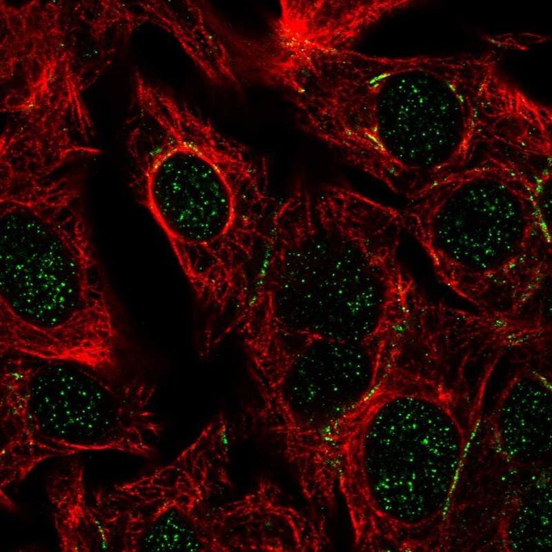
- Main image

- Experimental details
- Immunofluorescent staining of human cell line Hep G2 shows localization to nucleus & cell junctions.
- Sample type
- Human
Supportive validation
- Submitted by
- Atlas Antibodies (provider)
- Enhanced method
- Orthogonal validation
- Main image
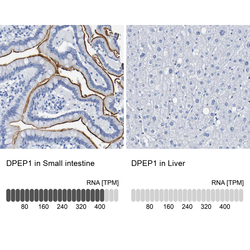
- Experimental details
- Immunohistochemistry analysis in human small intestine and liver tissues using HPA012783 antibody. Corresponding DPEP1 RNA-seq data are presented for the same tissues.
- Sample type
- Human
- Protocol
- Protocol
