 Explore
 Validate
 Learn
Learn Western blot
 Immunohistochemistry
Antibody data
- Antibody Data
- Antigen structure
- References [4]
- Comments [0]
- Validations
- Immunohistochemistry [3]
- Flow cytometry [1]
- Other assay [2]
Submit
Validation data
Reference
Comment
Report error
- Product number
- 44-1260G - Provider product page

- Provider
- Invitrogen Antibodies
- Product name
- Phospho-SGK1 (Thr256) Polyclonal Antibody
- Antibody type
- Polyclonal
- Antigen
- Synthetic peptide
- Reactivity
- Human, Mouse, Rat
- Host
- Rabbit
- Isotype
- IgG
- Vial size
- 100 μL
- Storage
- -20°C
Submitted references Cutaneous and hepatic vascular lesions due to a recurrent somatic GJA4 mutation reveal a pathway for vascular malformation.
Stimulation of Epithelial Sodium Channels in Endothelial Cells by Bone Morphogenetic Protein-4 Contributes to Salt-Sensitive Hypertension in Rats.
Glucocorticoids and serum- and glucocorticoid-inducible kinase 1 are potent regulators of CFTR in the native intestine: implications for stress-induced diarrhea.
The effects of brain serotonin deficiency on behavioural disinhibition and anxiety-like behaviour following mild early life stress.
Ugwu N, Atzmony L, Ellis KT, Panse G, Jain D, Ko CJ, Nassiri N, Choate KA
HGG advances 2021 Apr 8;2(2)
HGG advances 2021 Apr 8;2(2)
Stimulation of Epithelial Sodium Channels in Endothelial Cells by Bone Morphogenetic Protein-4 Contributes to Salt-Sensitive Hypertension in Rats.
Yang X, Niu N, Liang C, Wu MM, Tang LL, Wang QS, Lou J, Song BL, Zheng WW, Ma HP, Zhang ZR
Oxidative medicine and cellular longevity 2020;2020:3921897
Oxidative medicine and cellular longevity 2020;2020:3921897
Glucocorticoids and serum- and glucocorticoid-inducible kinase 1 are potent regulators of CFTR in the native intestine: implications for stress-induced diarrhea.
Ahsan MK, Figueroa-Hall L, Baratta V, Garcia-Milian R, Lam TT, Hoque K, Salas PJ, Ameen NA
American journal of physiology. Gastrointestinal and liver physiology 2020 Aug 1;319(2):G121-G132
American journal of physiology. Gastrointestinal and liver physiology 2020 Aug 1;319(2):G121-G132
The effects of brain serotonin deficiency on behavioural disinhibition and anxiety-like behaviour following mild early life stress.
Sachs BD, Rodriguiz RM, Siesser WB, Kenan A, Royer EL, Jacobsen JP, Wetsel WC, Caron MG
The international journal of neuropsychopharmacology 2013 Oct;16(9):2081-94
The international journal of neuropsychopharmacology 2013 Oct;16(9):2081-94
No comments: Submit comment
Supportive validation
- Submitted by
- Invitrogen Antibodies (provider)
- Main image
- Experimental details
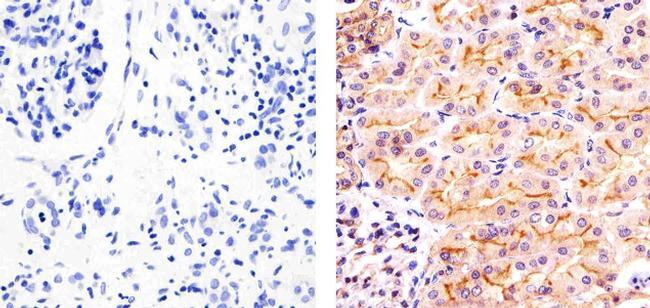
- Immunohistochemistry analysis of Phospho SGK1 (pT256) showing staining in the cytoplasm and membrane of paraffin-embedded human kidney tissue (right) compared to a negative control without primary antibody (left). To expose target proteins, antigen retrieval was performed using 10mM sodium citrate (pH 6.0), microwaved for 8-15 min. Following antigen retrieval, tissues were blocked in 3% H2O2-methanol for 15 min at room temperature, washed with ddH2O and PBS, and then probed with a Phospho SGK1 (pT256) Rabbit Polyclonal Antibody (Product # 44-1260G) diluted in 3% BSA-PBS at a dilution of 1:50 overnight at 4ºC in a humidified chamber. Tissues were washed extensively in PBST and detection was performed using an HRP-conjugated secondary antibody followed by colorimetric detection using a DAB kit. Tissues were counterstained with hematoxylin and dehydrated with ethanol and xylene to prep for mounting.
- Submitted by
- Invitrogen Antibodies (provider)
- Main image

- Experimental details
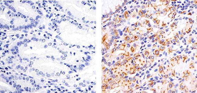
- Immunohistochemistry analysis of Phospho SGK1 (pT256) showing staining in the cytoplasm and membrane of paraffin-embedded human stomach tissue (right) compared to a negative control without primary antibody (left). To expose target proteins, antigen retrieval was performed using 10mM sodium citrate (pH 6.0), microwaved for 8-15 min. Following antigen retrieval, tissues were blocked in 3% H2O2-methanol for 15 min at room temperature, washed with ddH2O and PBS, and then probed with a Phospho SGK1 (pT256) Rabbit Polyclonal Antibody (Product # 44-1260G) diluted in 3% BSA-PBS at a dilution of 1:50 overnight at 4ºC in a humidified chamber. Tissues were washed extensively in PBST and detection was performed using an HRP-conjugated secondary antibody followed by colorimetric detection using a DAB kit. Tissues were counterstained with hematoxylin and dehydrated with ethanol and xylene to prep for mounting.
- Submitted by
- Invitrogen Antibodies (provider)
- Main image
- Experimental details
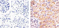
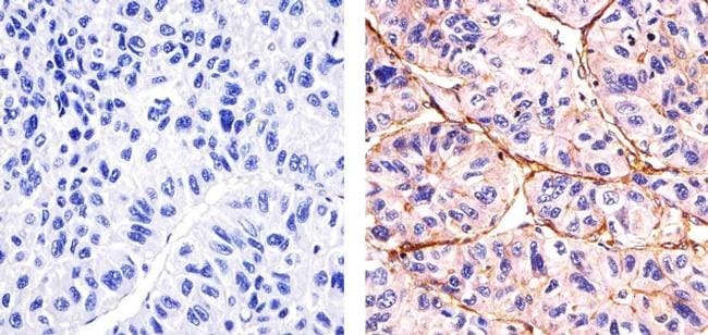
- Immunohistochemistry analysis of Phospho SGK1 (pT256) showing staining in the membrane and weakly in the cytoplasm of paraffin-embedded human hepatocarcinoma tissue (right) compared to a negative control without primary antibody (left). To expose target proteins, antigen retrieval was performed using 10mM sodium citrate (pH 6.0), microwaved for 8-15 min. Following antigen retrieval, tissues were blocked in 3% H2O2-methanol for 15 min at room temperature, washed with ddH2O and PBS, and then probed with a Phospho SGK1 (pT256) Rabbit Polyclonal Antibody (Product # 44-1260G) diluted in 3% BSA-PBS at a dilution of 1:20 overnight at 4ºC in a humidified chamber. Tissues were washed extensively in PBST and detection was performed using an HRP-conjugated secondary antibody followed by colorimetric detection using a DAB kit. Tissues were counterstained with hematoxylin and dehydrated with ethanol and xylene to prep for mounting.
Supportive validation
- Submitted by
- Invitrogen Antibodies (provider)
- Main image
- Experimental details
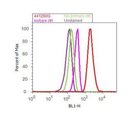
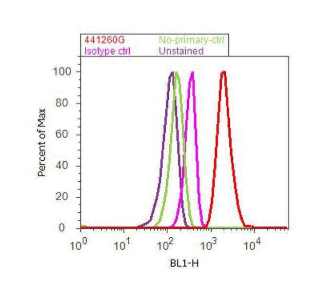
- Flow cytometry analysis of SGK1 [pThr256] was done on A549 cells treated with TGF beta (20ng/mL, 15 minutes). Cells were fixed with 70% ethanol for 10 minutes, permeabilized with 0.25% Triton™ X-100 for 20 minutes, and blocked with 5% BSA for 30 minutes at room temperature. Cells were labeled with SGK1 [pThr256] Rabbit Polyclonal Antibody (441260G, red histogram) or with rabbit isotype control (pink histogram) at 3-5 ug/million cells in 2.5% BSA. After incubation at room temperature for 2 hours, the cells were labeled with Alexa Fluor® 488 Goat Anti-Rabbit Secondary Antibody (A11008) at a dilution of 1:400 for 30 minutes at room temperature. The representative 10,000 cells were acquired and analyzed for each sample using an Attune® Acoustic Focusing Cytometer. The purple histogram represents unstained control cells and the green histogram represents no-primary-antibody control.
Supportive validation
- Submitted by
- Invitrogen Antibodies (provider)
- Main image
- Experimental details
- NULL

- Submitted by
- Invitrogen Antibodies (provider)
- Main image
- Experimental details
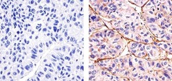
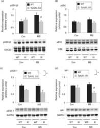
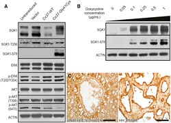
- Figure 3. Cx37-Gly41Cys expression activates SGK1 via a non-canonical pathway (A) HUVECs transduced with Cx37-G41C express a higher molecular weight phosphorylated form of SGK1, with expression of SGK1-Ser78 seen exclusively in Cx37-Gly41Cys cells. Cx37-Gly41Cys does not alter MAPK expression. Cx37-WT leads to decreased Akt-Ser473 expression, whereas Cx37-Gly41Cys expression results in almost complete abrogation. Actin is blotted as a loading control. (B) Gradient induction of Cx37-Gly41Cys expression with doxycycline leads to dose-dependent increases in phosphorylation of SGK1 at serine 78 and an increase in total SGK1 expression. In the absence of Cx37-Gly41Cys expression, SGK1 runs as a doublet on SDS-PAGE (lane 1) but migrates as a single band when expression of the mutant protein is induced. These data indicate that Cx37-Gly41Cys leads to SGK1 activation via serine 78 phosphorylation. Actin is blotted as a loading control. (C) Immunohistochemical staining reveals absence of SGK1-Ser78 in vasculature of normal liver tissue and strong SGK1-Ser78 expression in HH tissue. Scale bars, 75 mum.
