Explore
 Validate
 Learn
Learn Western blot
 Immunohistochemistry
Antibody data
- Antibody Data
- Antigen structure
- References [1]
- Comments [0]
- Validations
- Immunohistochemistry [1]
Submit
Validation data
Reference
Comment
Report error
- Product number
- HPA019639 - Provider product page

- Provider
- Atlas Antibodies
- Proper citation
- Atlas Antibodies Cat#HPA019639, RRID:AB_1845970
- Product name
- Anti-CANT1
- Antibody type
- Polyclonal
- Description
- Polyclonal Antibody against Human CANT1, Gene description: calcium activated nucleotidase 1, Alternative Gene Names: SCAN-1, SHAPY, Validated applications: WB, IHC, Uniprot ID: Q8WVQ1, Storage: Store at +4°C for short term storage. Long time storage is recommended at -20°C.
- Reactivity
- Human
- Host
- Rabbit
- Conjugate
- Unconjugated
- Isotype
- IgG
- Vial size
- 100 µl
- Concentration
- 0.2 mg/ml
- Storage
- Store at +4°C for short term storage. Long time storage is recommended at -20°C.
- Handling
- The antibody solution should be gently mixed before use.
Submitted references Initial Quantitative Proteomic Map of 28 Mouse Tissues Using the SILAC Mouse
Geiger T, Velic A, Macek B, Lundberg E, Kampf C, Nagaraj N, Uhlen M, Cox J, Mann M
Molecular & Cellular Proteomics 2013;12(6):1709-1722
Molecular & Cellular Proteomics 2013;12(6):1709-1722
No comments: Submit comment
Supportive validation
- Submitted by
- Atlas Antibodies (provider)
- Enhanced method
- Orthogonal validation
- Main image
- Experimental details
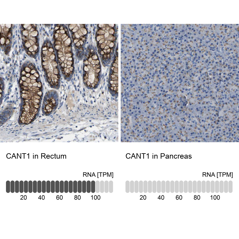
- Immunohistochemistry analysis in human rectum and pancreas tissues using Anti-CANT1 antibody. Corresponding CANT1 RNA-seq data are presented for the same tissues.
- Sample type
- Human
- Protocol
- Protocol