Explore
 Validate
 Learn
Learn Western blot
Antibody data
- Antibody Data
- Antigen structure
- References [0]
- Comments [0]
- Validations
- Western blot [4]
- Immunohistochemistry [1]
Submit
Validation data
Reference
Comment
Report error
- Product number
- GTX105682 - Provider product page

- Provider
- GeneTex
- Proper citation
- GeneTex Cat#GTX105682, RRID:AB_1950977
- Product name
- NCKAP1 antibody [C1C2], Internal
- Antibody type
- Polyclonal
- Reactivity
- Human, Mouse, Rat
- Host
- Rabbit
No comments: Submit comment
Supportive validation
- Submitted by
- GeneTex (provider)
- Main image
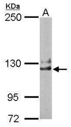
- Experimental details
- Sample (50 ug of whole cell lysate) A: Mouse brain 5% SDS PAGE GTX105682 diluted at 1:1000

- Submitted by
- GeneTex (provider)
- Main image

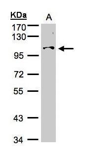
- Experimental details
- Sample (30£gg whole cell lysate)A:Hep G2 (GTX27900)7.5% SDS PAGEGTX105682 diluted at 1:500
- Submitted by
- GeneTex (provider)
- Main image

- Experimental details
- NCKAP1 antibody [C1C2], Internal detects NCKAP1 protein by Western blot analysis.A. 50 ?g rat brain lysate/extract5 % SDS-PAGENCKAP1 antibody [C1C2], Internal (GTX105682) dilution: 1:1000

- Submitted by
- GeneTex (provider)
- Main image
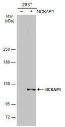
- Experimental details
- Non-transfected (¡V) and transfected (+) 293T whole cell extracts (30 ?g) were separated by 5% SDS-PAGE, and the membrane was blotted with NCKAP1 antibody [C1C2], Internal (GTX105682) diluted at 1:5000. The HRP-conjugated anti-rabbit IgG antibody (GTX213110-01) was used to detect the primary antibody.

Supportive validation
- Submitted by
- GeneTex (provider)
- Main image
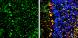
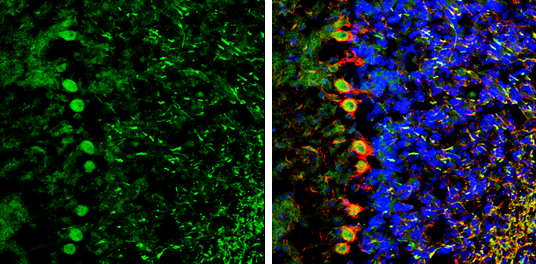
- Experimental details
- NCKAP1 antibody [C1C2], Internal detects NCKAP1 protein by immunohistochemical analysis.Sample: Frozen-sectioned mouse cerebellum.Green: NCKAP1 stained by NCKAP1 antibody [C1C2], Internal (GTX105682) diluted at 1:250.Red: NF-H, stained by NF-H antibody [GT114] (GTX634289) diluted at 1:500.Blue: Fluoroshield with DAPI (GTX30920).