 Explore
 Validate
 Learn
Learn Western blot
Antibody data
- Antibody Data
- Antigen structure
- References [0]
- Comments [0]
- Validations
- Western blot [2]
- Immunohistochemistry [1]
Submit
Validation data
Reference
Comment
Report error
- Product number
- ACC-083-200UL - Provider product page

- Provider
- Invitrogen Antibodies
- Product name
- TRPML3 (Mucolipin 3) Polyclonal Antibody
- Antibody type
- Polyclonal
- Antigen
- Other
- Reactivity
- Human, Mouse, Rat
- Host
- Rabbit
- Isotype
- IgG
- Vial size
- 200 µL
- Concentration
- 0.8 mg/mL
- Storage
- -20° C, Avoid Freeze/Thaw Cycles
No comments: Submit comment
Supportive validation
- Submitted by
- Invitrogen Antibodies (provider)
- Main image

- Experimental details
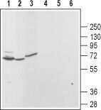
- Western blot analysis of rat brain (lanes 1 and 4), kidney (lanes 2 and 5) and pancreas (lanes 3 and 6): - 1,2,3. Anti-TRPML3 (Mucolipin 3) Antibody (#ACC-083), (1:200).4,5,6. Anti-TRPML3 (Mucolipin 3) Antibody , preincubated with TRPML3/Mucolipin 3 Blocking Peptide (#BLP-CC083).
- Submitted by
- Invitrogen Antibodies (provider)
- Main image

- Experimental details
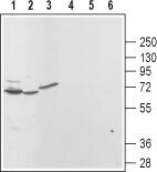
- Western blot analysis of rat brain (lanes 1 and 4), kidney (lanes 2 and 5) and pancreas (lanes 3 and 6): - 1,2,3. Anti-TRPML3 (Mucolipin 3) Antibody (#ACC-083), (1:200).4,5,6. Anti-TRPML3 (Mucolipin 3) Antibody , preincubated with TRPML3/Mucolipin 3 Blocking Peptide (#BLP-CC083).
Supportive validation
- Submitted by
- Invitrogen Antibodies (provider)
- Main image
- Experimental details
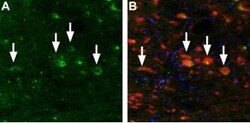
- Expression of Mucolipin 3 in mouse brain stem - Immunohistochemical staining of TRPML3 in mouse brain stem using Anti-TRPML3 (Mucolipin 3) Antibody (#ACC-083). A. TRPML3 (green) appears in neurons (arrows) in the area of the dorsal cochlear nucleus. B. Staining of the same section with mouse Anti-parvalbumin (red) reveals that the TRPML3 appears in a subset of neurons in this nucleus. DAPI is used as the counterstain (blue).
