 Explore
 Validate
 Learn
Learn Western blot
Antibody data
- Antibody Data
- Antigen structure
- References [3]
- Comments [0]
- Validations
- Western blot [1]
Submit
Validation data
Reference
Comment
Report error
- Product number
- MAB2043 - Provider product page

- Provider
- Abnova Corporation
- Proper citation
- Abnova Corporation Cat#MAB2043, RRID:AB_1672357
- Product name
- CYR61 monoclonal antibody, clone 3H3
- Antibody type
- Monoclonal
- Description
- Mouse monoclonal antibody raised against partial recombinant CYR61.
- Isotype
- IgG
- Antibody clone number
- 3H3
- Storage
- Store at -20°C.Aliquot to avoid repeated freezing and thawing.
Submitted references Cyr61 suppresses the growth of non-small-cell lung cancer cells via the beta-catenin-c-myc-p53 pathway.
Xenopus Cyr61 regulates gastrulation movements and modulates Wnt signalling.
CYR61 stimulates human skin fibroblast migration through Integrin alpha vbeta 5 and enhances mitogenesis through integrin alpha vbeta 3, independent of its carboxyl-terminal domain.
Tong X, O'Kelly J, Xie D, Mori A, Lemp N, McKenna R, Miller CW, Koeffler HP
Oncogene 2004 Jun 17;23(28):4847-55
Oncogene 2004 Jun 17;23(28):4847-55
Xenopus Cyr61 regulates gastrulation movements and modulates Wnt signalling.
Latinkic BV, Mercurio S, Bennett B, Hirst EM, Xu Q, Lau LF, Mohun TJ, Smith JC
Development (Cambridge, England) 2003 Jun;130(11):2429-41
Development (Cambridge, England) 2003 Jun;130(11):2429-41
CYR61 stimulates human skin fibroblast migration through Integrin alpha vbeta 5 and enhances mitogenesis through integrin alpha vbeta 3, independent of its carboxyl-terminal domain.
Grzeszkiewicz TM, Kirschling DJ, Chen N, Lau LF
The Journal of biological chemistry 2001 Jun 15;276(24):21943-50
The Journal of biological chemistry 2001 Jun 15;276(24):21943-50
No comments: Submit comment
Supportive validation
- Submitted by
- Abnova Corporation (provider)
- Main image
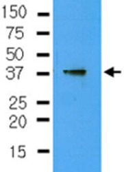
- Experimental details
- The cell lysate of Jurkat (40 ug) was resolved by SDS-PAGE and probed with CYR61 monoclonal antibody, clone 3H3 (Cat # MAB2043) (1: 250). Proteins were visualized using a goat anti-mouse secondary antibody conjugated to HRP and an ECL detection system.
