Explore
 Validate
 Learn
Learn Western blot
 Immunocytochemistry
Antibody data
- Antibody Data
- Antigen structure
- References [1]
- Comments [0]
- Validations
- Immunocytochemistry [3]
- Immunohistochemistry [3]
Submit
Validation data
Reference
Comment
Report error
- Product number
- PA5-78022 - Provider product page

- Provider
- Invitrogen Antibodies
- Product name
- CYR61 Polyclonal Antibody
- Antibody type
- Polyclonal
- Antigen
- Recombinant full-length protein
- Description
- Positive Control: HeLa (1X Brefeldin A for 24hr) Predicted Reactivity: Chicken (81%), Chimpanzee (99%), Bovine (92%) Store product as a concentrated solution. Centrifuge briefly prior to opening the vial.
- Reactivity
- Human, Mouse, Rat
- Host
- Rabbit
- Isotype
- IgG
- Vial size
- 100 μL
- Concentration
- 1.34 mg/mL
- Storage
- Store at 4°C short term. For long term storage, store at -20°C, avoiding freeze/thaw cycles.
Submitted references Cul4A-DDB1-mediated monoubiquitination of phosphoglycerate dehydrogenase promotes colorectal cancer metastasis via increased S-adenosylmethionine.
Zhang Y, Yu H, Zhang J, Gao H, Wang S, Li S, Wei P, Liang J, Yu G, Wang X, Li X, Li D, Yang W
The Journal of clinical investigation 2021 Nov 1;131(21)
The Journal of clinical investigation 2021 Nov 1;131(21)
No comments: Submit comment
Supportive validation
- Submitted by
- Invitrogen Antibodies (provider)
- Main image

- Experimental details
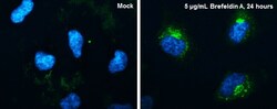
- Immunofluorescent analysis of CYR61 in HeLa cells. Samples were treated with 4% paraformaldehyde at RT for 15 min and incubated with CYR61 polyclonal antibody (Product # PA5-78022) using a dilution of 1:200, followed by Hoechst.
- Submitted by
- Invitrogen Antibodies (provider)
- Main image

- Experimental details
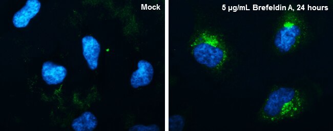
- CYR61 Polyclonal Antibody detects CYR61 protein at endoplasmic reticulum by immunofluorescent analysis. Sample: HeLa cells were fixed in 4% paraformaldehyde at RT for 15 min. Green: CYR61 protein stained by CYR61 Polyclonal Antibody (Product # PA5-78022) diluted at 1:200. Blue: Hoechst 33342 staining.
- Submitted by
- Invitrogen Antibodies (provider)
- Main image
- Experimental details
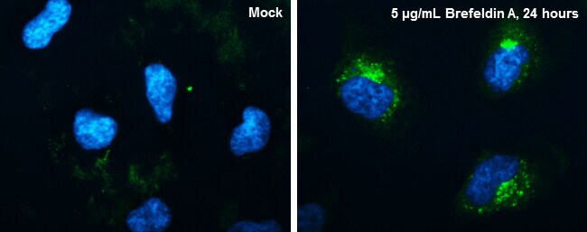
- CYR61 Polyclonal Antibody detects CYR61 protein at endoplasmic reticulum by immunofluorescent analysis. Sample: HeLa cells were fixed in 4% paraformaldehyde at RT for 15 min. Green: CYR61 protein stained by CYR61 Polyclonal Antibody (Product # PA5-78022) diluted at 1:200. Blue: Hoechst 33342 staining.

Supportive validation
- Submitted by
- Invitrogen Antibodies (provider)
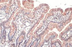
- Main image
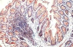
- Experimental details
- CYR61 Polyclonal Antibody detects secreted CYR61 protein by immunohistochemical analysis. Sample: Paraffin-embedded mouse colon. CYR61 stained by CYR61 Polyclonal Antibody (Product # PA5-78022) diluted at 1:500. Antigen Retrieval: Citrate buffer, pH 6.0, 15 min.
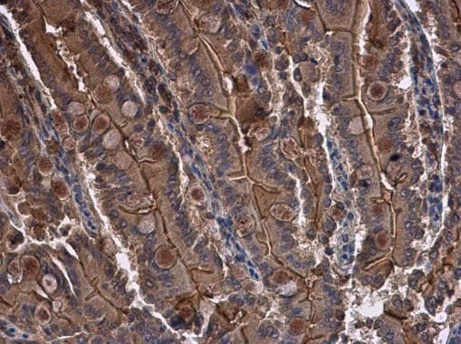
- Submitted by
- Invitrogen Antibodies (provider)
- Main image

- Experimental details
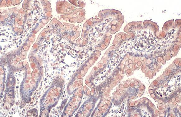
- CYR61 Polyclonal Antibody detects CYR61 protein at cytoplasm in rat intestine by immunohistochemical analysis. Sample: Paraffin-embedded rat intestine. CYR61 Polyclonal Antibody (Product # PA5-78022) diluted at 1:500. Antigen Retrieval: Citrate buffer, pH 6.0, 15 min.
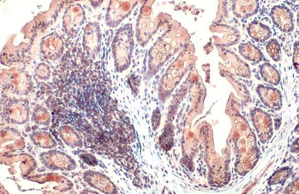
- Submitted by
- Invitrogen Antibodies (provider)
- Main image
- Experimental details
- CYR61 Polyclonal Antibody detects secreted CYR61 protein by immunohistochemical analysis. Sample: Paraffin-embedded rat colon. CYR61 stained by CYR61 Polyclonal Antibody (Product # PA5-78022) diluted at 1:500. Antigen Retrieval: Citrate buffer, pH 6.0, 15 min.