Explore
 Validate
 Learn
Learn Western blot
Antibody data
- Antibody Data
- Antigen structure
- References [1]
- Comments [0]
- Validations
- Western blot [2]
- Immunohistochemistry [1]
- Other assay [1]
Submit
Validation data
Reference
Comment
Report error
- Product number
- PA1-16579 - Provider product page

- Provider
- Invitrogen Antibodies
- Product name
- CYR61 Polyclonal Antibody
- Antibody type
- Polyclonal
- Antigen
- Synthetic peptide
- Description
- This antibody is predicted to react with rat and mouse based on 88% sequence homology. Suggested positive control: antigen standard for CYR61 (transient overexpression lysate), MDA-MB-231 breast cancer cell lysate.
- Reactivity
- Human, Mouse, Rabbit
- Host
- Rabbit
- Isotype
- IgG
- Vial size
- 100 μL
- Concentration
- 1 mg/mL
- Storage
- Store at 4°C short term. For long term storage, store at -20°C, avoiding freeze/thaw cycles.
Submitted references Aberrant activation of CYR61 enhancers in colorectal cancer development.
Xie L, Song X, Lin H, Chen Z, Li Q, Guo T, Xu T, Su T, Xu M, Chang X, Wang LK, Liang B, Huang D
Journal of experimental & clinical cancer research : CR 2019 May 22;38(1):213
Journal of experimental & clinical cancer research : CR 2019 May 22;38(1):213
No comments: Submit comment
Supportive validation
- Submitted by
- Invitrogen Antibodies (provider)
- Main image
- Experimental details
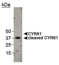
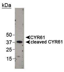
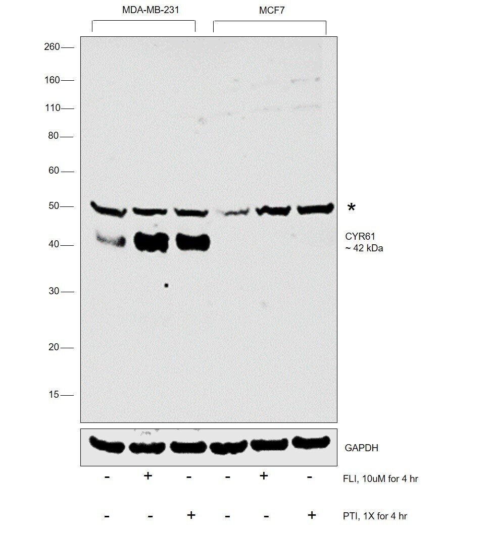
- Western Blot detection of cleaved CYR61 in MDA-MB-231 cell lysate using Product # PA1-16579.
- Submitted by
- Invitrogen Antibodies (provider)
- Main image
- Experimental details
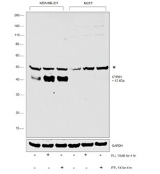
- Western blot was performed using Anti- CYR61 Polyclonal Antibody (Product # PA1-16579) and a ~42 kDa band corresponding to CYR61 was increased upon treating the cells with secretion blockers (FLI and PTI) in MDA-MB-231 when compared to MCF7. An uncharacterized band (*) was observed at ~ 50 kDa. Whole cell extracts (30 µg lysate) of MDA-MB-231 (Lane 1), MDA-MB-231 treated with FLI (10µM for 4 hr) (Lane 2), MDA-MB-231 treated with PTI (1X for 4 hr) (Lane 3), MCF7 (Lane 4), MCF7 treated with FLI (10µM for 4 hr) (Lane 5) and MCF7 treated with PTI (1X for 4 hr) (Lane 6) were electrophoresed using Novex® NuPAGE® 4-12 % Bis-Tris gel (Product # NP0321BOX). Resolved proteins were then transferred onto a nitrocellulose membrane (Product # IB23001) by iBlot® 2 Dry Blotting System (Product # IB21001). The blot was probed with the primary antibody (1:500 dilution) and detected by chemiluminescence with Goat anti-Rabbit IgG (Heavy Chain) Superclonal™ Recombinant Secondary Antibody, HRP (Product # A27036, 1:4000 dilution) using the iBright FL 1000 (Product # A32752). Chemiluminescent detection was performed using Novex® ECL Chemiluminescent Substrate Reagent Kit (Product # WP20005).
Supportive validation
- Submitted by
- Invitrogen Antibodies (provider)
- Main image
- Experimental details
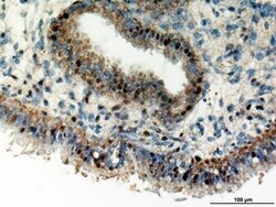
- Immunohistochemical staining of Human endometrium using Product # PA1-16579.

Supportive validation
- Submitted by
- Invitrogen Antibodies (provider)
- Main image
- Experimental details
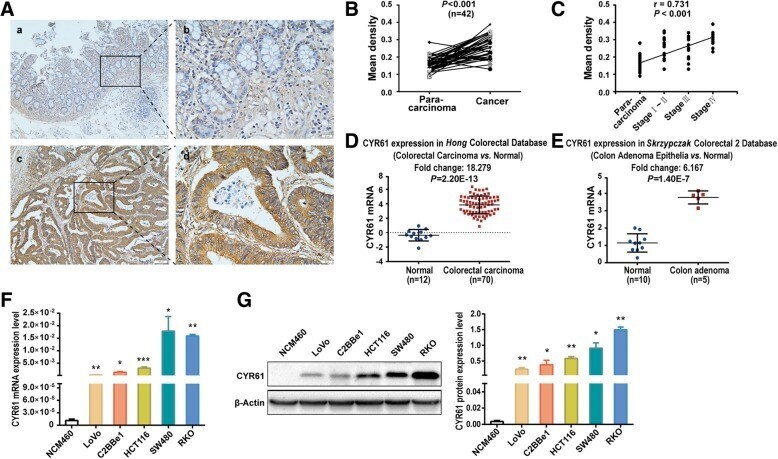
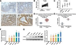
- Fig. 1 CYR61 expression levels in primary colonic adenocarcinoma patient samples and colon cell lines. a Immunostaining for CYR61 in paraffin sections of para-carcinoma tissues ( a , b and cancer tissues ( c , d from patients with colon adenocarcinoma (magnification, x 100, x 400). b The mean density of CYR61 in IHC of colon para-carcinoma and cancer tissues from 42 cases; significance determined by the paired-samples t-test. c Correlation between the mean density of CYR61 in IHC and TNM levels, significance determined by Spearman's rank correlation. d CYR61 mRNA expression levels in primary colon cancer tissues compared with normal controls by analysis of the Hong Colorectal microarray dataset. e CYR61 mRNA expression levels in primary colon cancer tissues compared with normal controls by analysis of the Skrzypczak Colorectal 2 microarray dataset. f Expression of CYR61 mRNA and g protein levels in human colon cell lines, significance determined by the independent samples t-test. Data are shown as mean +- S.D., n = 3. * P < 0.05, ** P < 0.01, *** P < 0.001