Explore
 Validate
 Learn
Learn Western blot
Antibody data
- Antibody Data
- Antigen structure
- References [0]
- Comments [0]
- Validations
- Western blot [2]
- Immunocytochemistry [1]
- Immunohistochemistry [1]
Submit
Validation data
Reference
Comment
Report error
- Product number
- TA328625 - Provider product page

- Provider
- OriGene
- Product name
- Rabbit Polyclonal Anti-Bombesin Receptor 1
- Antibody type
- Polyclonal
- Description
- Rabbit Polyclonal Anti-Bombesin Receptor 1
- Host
- Rabbit
- Conjugate
- Unconjugated
- Epitope
- NMBR
- Antibody clone number
- NULL
- Vial size
- 200 µl
- Concentration
- NULL
No comments: Submit comment
Supportive validation
- Submitted by
- OriGene (provider)
- Main image
- Experimental details
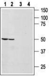
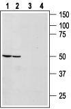
- Western blot analysis of rat (lanes 1 and 3) and mouse (lanes 2 and 4) brain lysates: 1, 2. Anti-Bombesin Receptor 1 antibody, (1:1000). 3, 4. Anti-Bombesin Receptor 1 antibody, preincubated with the control peptide antigen.
- Validation comment
- WB
- Submitted by
- OriGene (provider)
- Main image
- Experimental details
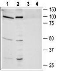
- Western blot analysis of human lung carcinoma NCI-H526 (lanes 1 and 3) and human prostate carcinoma PC-3 (lanes 2 and 4) cell line lysates: 1, 2. Anti-Bombesin Receptor 1 antibody, (1:1000). 3, 4. Anti-Bombesin Receptor 1 antibody, preincubated with the control peptide antigen.
- Validation comment
- WB

Supportive validation
- Submitted by
- OriGene (provider)
- Main image
- Experimental details
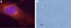
- Expression of Bombesin Receptor 1 in a human breast cancer cell line. Immunocytochemical staining of paraformaldehyde-fixed and permeabilized mammary gland adenocarcinoma MDA-MB-231. A. Cells were stained with Anti-Bombesin Receptor 1 antibody, (1:1000), followed by goat anti-rabbit-AlexaFluor-555 secondary antibody (red). Hoechst 33342 (blue) is used to visulaize the nuclei. B. Live view of the same field as in (A).
- Validation comment
- IF

Supportive validation
- Submitted by
- OriGene (provider)
- Main image
- Experimental details
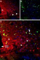
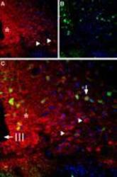
- Expression of Bombesin Receptor 1 in rat hypothalamus. Immunohistochemical staining of frozen rat hypothalamus sections using Anti-Bombesin Receptor 1 antibody , (1:200). A. Staining (red) appears in the neuropil near the ventricle (asterisk) and in neurons (triangles). Calbindin D28k staining (green) appears in neurons. Merge of (A) and (B) shows co-expression of Bombesin Receptor 1 and Calbindin in a few neurons (vertical arrow). DAPI is used as the counterstain.
- Validation comment
- IHC