Explore
 Validate
 Learn
Learn Western blot
 Immunocytochemistry
Antibody data
- Antibody Data
- Antigen structure
- References [0]
- Comments [0]
- Validations
- Western blot [6]
- Immunohistochemistry [2]
Submit
Validation data
Reference
Comment
Report error
- Product number
- NBP1-32622 - Provider product page

- Provider
- Novus Biologicals
- Proper citation
- Novus Cat#NBP1-32622, RRID:AB_2114358
- Product name
- Rabbit Polyclonal TFIIB Antibody
- Antibody type
- Polyclonal
- Description
- Immunogen affinity purified.
- Reactivity
- Human
- Host
- Rabbit
- Isotype
- IgG
- Vial size
- 100 ul
- Storage
- Aliquot and store at -20C or -80C. Avoid freeze-thaw cycles.
No comments: Submit comment
Supportive validation
- Submitted by
- Novus Biologicals (provider)
- Main image
- Experimental details
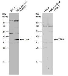
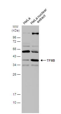
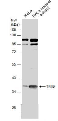
- Western Blot: TFIIB Antibody [NBP1-32622] - HeLa whole cell and nuclear extracts (30 ug) were separated by 12% SDS-PAGE, and the membranes were blotted with TFIIB antibody [N1C3]. The HRP-conjugated anti-rabbit IgG antibody was used to detect the primary antibody.
- Submitted by
- Novus Biologicals (provider)
- Main image
- Experimental details
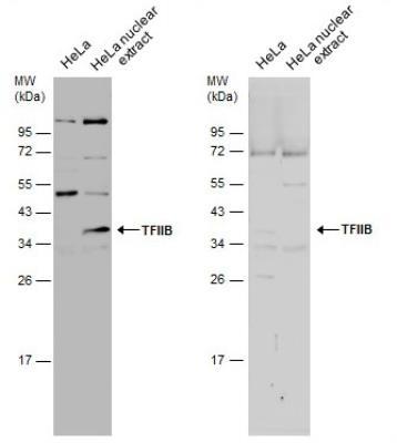
- Western Blot: TFIIB Antibody [NBP1-32622] - HeLa whole cell and nuclear extracts (30 ug) were separated by 10% SDS-PAGE, and the membrane was blotted with TFIIB antibody [N1C3] diluted at 1:1500. The HRP-conjugated anti-rabbit IgG antibody (NBP2-19301) was used to detect the primary antibody.
- Submitted by
- Novus Biologicals (provider)
- Main image
- Experimental details
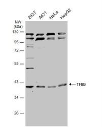
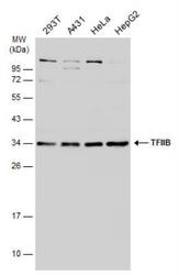
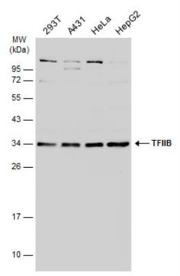
- Western Blot: TFIIB Antibody [NBP1-32622] - Various whole cell extracts (30 ug) were separated by 10% SDS-PAGE, and the membrane was blotted with TFIIB antibody [N1C3] diluted at 1:500. The HRP-conjugated anti-rabbit IgG antibody (NBP2-19301) was used to detect the primary antibody.
- Submitted by
- Novus Biologicals (provider)
- Main image
- Experimental details
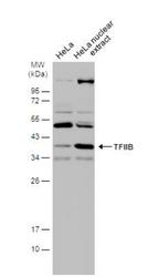
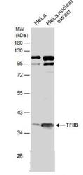
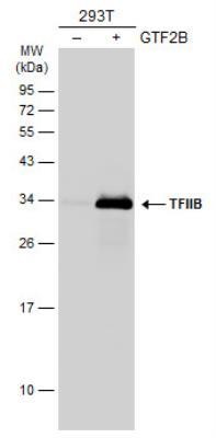
- Western Blot: TFIIB Antibody [NBP1-32622] - HeLa whole cell and nuclear extracts (30 ug) were separated by 10% SDS-PAGE, and the membranes were blotted with TFIIB antibody [N1C3] (diluted at 1:500.
- Submitted by
- Novus Biologicals (provider)
- Main image
- Experimental details
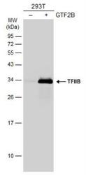
- Western Blot: TFIIB Antibody [NBP1-32622] - Non-transfected (-) and transfected (+) 293T whole cell extracts (30 ug) were separated by 12% SDS-PAGE, and the membrane was blotted with TFIIB antibody diluted at 1:2000. HRP-conjugated anti-rabbit IgG antibody was used to detect the primary antibody.
- Submitted by
- Novus Biologicals (provider)
- Main image
- Experimental details
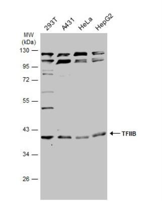
- Western Blot: TFIIB Antibody [NBP1-32622] - Various whole cell extracts (30 ug) were separated by 12% SDS-PAGE, and the membrane was blotted with TFIIB antibody diluted at 1:1000. HRP-conjugated anti-rabbit IgG antibody was used to detect the primary antibody.
Supportive validation
- Submitted by
- Novus Biologicals (provider)
- Main image

- Experimental details
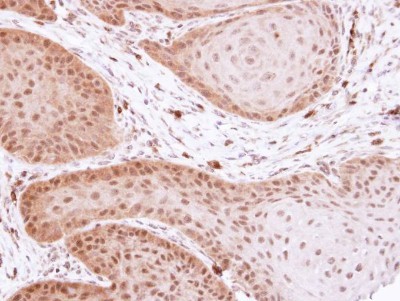
- Immunohistochemistry-Paraffin: TFIIB Antibody [NBP1-32622] - Paraffin-embedded Cal27 xenograft, using antibody at 1:100 dilution.
- Submitted by
- Novus Biologicals (provider)
- Main image
- Experimental details
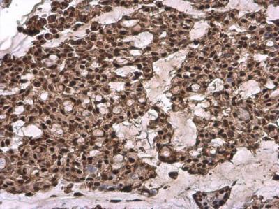
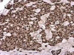
- Immunohistochemistry-Paraffin: TFIIB Antibody [NBP1-32622] - TFIIB antibody detects TFIIB protein at cytoplasm and nucleus by immunohistochemical analysis. Sample: Paraffin-embedded human breast carcinoma. TFIIB stained by TFIIB antibody diluted at 1:500.