Explore
 Validate
 Learn
Learn Western blot
Antibody data
- Antibody Data
- Antigen structure
- References [1]
- Comments [0]
- Validations
- Western blot [1]
- Immunocytochemistry [3]
Submit
Validation data
Reference
Comment
Report error
- Product number
- PA3-840 - Provider product page

- Provider
- Invitrogen Antibodies
- Product name
- mDIC Polyclonal Antibody
- Antibody type
- Polyclonal
- Antigen
- Synthetic peptide
- Description
- PA3-840 detects mitochondrial dicarboxylate carrier protein(mDIC) in human and mouse cells. PA3-840 has been successfully used in Western blotting and ICC/IF procedures. By Western blot it detects a 28 kDa protein representing mDIC. The PA3-840 immunogen is a synthetic peptide corresponding to the C-terminal residues L(276) R K H F G I K V P T T(287) of mDIC.
- Reactivity
- Human, Mouse
- Host
- Rabbit
- Isotype
- IgG
- Vial size
- 100 µL
- Concentration
- Conc. Not Determined
- Storage
- -20° C, Avoid Freeze/Thaw Cycles
Submitted references Predominant expression of the mitochondrial dicarboxylate carrier in white adipose tissue.
Das K, Lewis RY, Combatsiaris TP, Lin Y, Shapiro L, Charron MJ, Scherer PE
The Biochemical journal 1999 Dec 1;344 Pt 2(Pt 2):313-20
The Biochemical journal 1999 Dec 1;344 Pt 2(Pt 2):313-20
No comments: Submit comment
Supportive validation
- Submitted by
- Invitrogen Antibodies (provider)
- Main image
- Experimental details
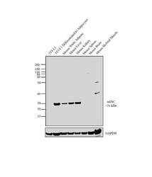
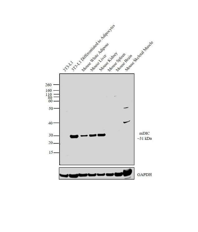
- Western blot analysis was performed on membrane enriched extracts (30 µg lysate) of 3T3-L1 (Lane 1), 3T3-L1 differentiated to adipocytes (Lane 2), tissue extracts of Mouse White Adipose (Lane 3), Mouse Liver (Lane 4), Mouse Kidney (Lane 5), Mouse Spleen (Lane 6), Mouse Brain (Lane7) and Mouse Skeletal Muscle (Lane 8). The blot was probed with mDIC Polyclonal Antibody (Product # PA3-840, 1:500 dilution) and detected by chemiluminescence using Goat anti-Rabbit IgG (H+L) Superclonal™ Secondary Antibody, HRP conjugate (Product # A27036, 0.25 µg/mL, 1:4000 dilution). A 31 kDa band corresponding to mDIC was observed in Mouse White Adipose, Mouse Liver, Mouse Kidney compared to other tissue extracts tested and was also enhanced in 3T3-L1 after differentiating it to adipocytes.
Supportive validation
- Submitted by
- Invitrogen Antibodies (provider)
- Main image
- Experimental details
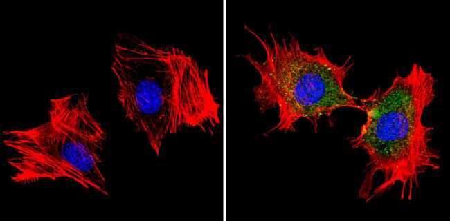
- Immunofluorescent analysis of MDC1 (green) showing staining in the cytoplasm of 3T3-L1 cells (right) compared to a negative control without primary antibody (left). Formalin-fixed cells were permeabilized with 0.1% Triton X-100 in TBS for 5-10 minutes and blocked with 3% BSA-PBS for 30 minutes at room temperature. Cells were probed with a MDC1 polyclonal antibody (Product # PA3-840) in 3% BSA-PBS at a dilution of 1:100 and incubated overnight at 4ºC in a humidified chamber. Cells were washed with PBST and incubated with a DyLight-conjugated secondary antibody in PBS at room temperature in the dark. Actin was stained using Alexa Fluor 554 (red) and nuclei were stained with Hoechst or DAPI (blue). Images were taken at a magnification of 60x.
- Submitted by
- Invitrogen Antibodies (provider)
- Main image

- Experimental details
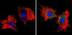
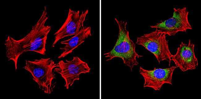
- Immunofluorescent analysis of MDC1 (green) showing staining in the cytoplasm and nucleus of C2C12 cells (right) compared to a negative control without primary antibody (left). Formalin-fixed cells were permeabilized with 0.1% Triton X-100 in TBS for 5-10 minutes and blocked with 3% BSA-PBS for 30 minutes at room temperature. Cells were probed with a MDC1 polyclonal antibody (Product # PA3-840) in 3% BSA-PBS at a dilution of 1:100 and incubated overnight at 4ºC in a humidified chamber. Cells were washed with PBST and incubated with a DyLight-conjugated secondary antibody in PBS at room temperature in the dark. Actin was stained using Alexa Fluor 554 (red) and nuclei were stained with Hoechst or DAPI (blue). Images were taken at a magnification of 60x.
- Submitted by
- Invitrogen Antibodies (provider)
- Main image
- Experimental details
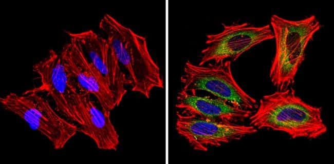
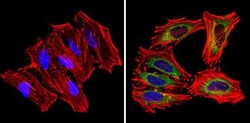
- Immunofluorescent analysis of MDC1 (green) showing staining in the cytoplasm of Hela cells (right) compared to a negative control without primary antibody (left). Formalin-fixed cells were permeabilized with 0.1% Triton X-100 in TBS for 5-10 minutes and blocked with 3% BSA-PBS for 30 minutes at room temperature. Cells were probed with a MDC1 polyclonal antibody (Product # PA3-840) in 3% BSA-PBS at a dilution of 1:100 and incubated overnight at 4ºC in a humidified chamber. Cells were washed with PBST and incubated with a DyLight-conjugated secondary antibody in PBS at room temperature in the dark. Actin was stained using Alexa Fluor 554 (red) and nuclei were stained with Hoechst or DAPI (blue). Images were taken at a magnification of 60x.