Explore
 Validate
 Learn
Learn Western blot
 Immunocytochemistry
Antibody data
- Antibody Data
- Antigen structure
- References [0]
- Comments [0]
- Validations
- Immunocytochemistry [2]
Submit
Validation data
Reference
Comment
Report error
- Product number
- PA1-545 - Provider product page

- Provider
- Invitrogen Antibodies
- Product name
- PCYT2 Polyclonal Antibody
- Antibody type
- Polyclonal
- Antigen
- Synthetic peptide
- Description
- PA1-545 detects ethanolaminephosphate cytidyltransferase (ECT) protein in human, rat, mouse and non-human primate samples. PA1-545 has successfully been used in Western blot procedures. By Western blot, this antibody detects an ~50 kDa protein representing ECT from C3H/10T1/2 cell lysates. PA1-545 immunizing peptide corresponds to amino acid residues 157-172 from human ECT protein. This immunizing peptide (PEP-241) can be used for neutralization and control experiments.
- Reactivity
- Human, Mouse, Rat
- Host
- Rabbit
- Isotype
- IgG
- Vial size
- 100 μg
- Concentration
- 0.4 mg/mL
- Storage
- -20°C, Avoid Freeze/Thaw Cycles
No comments: Submit comment
Supportive validation
- Submitted by
- Invitrogen Antibodies (provider)
- Main image
- Experimental details
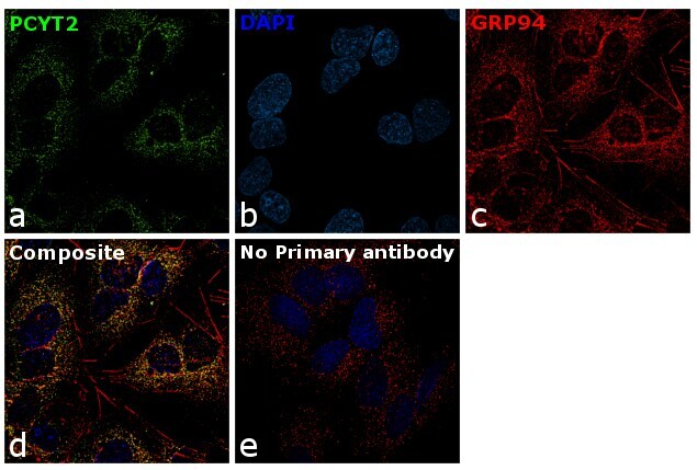
- Immunofluorescence analysis of PCYT2 was performed using 70% confluent log phase Hep G2 cells. The cells were fixed with 4% paraformaldehyde for 10 minutes, permeabilized with 0.1% Triton™ X-100 for 15 minutes, and blocked with 1% BSA for 1 hour at room temperature. The cells were labeled with PCYT2 Polyclonal Antibody (Product # PA1-545) at 5 µg/mL in 0.1% BSA, incubated at 4 degree Celsius overnight and then labeled with Goat anti-Rabbit IgG (H+L) Superclonal™ Secondary Antibody, Alexa Fluor® 488 conjugate (Product # A27034) at a dilution of 1:2000 for 45 minutes at room temperature (Panel a: green). Nuclei (Panel b: blue) were stained with ProLong™ Diamond Antifade Mountant with DAPI (Product # P36962). GRP94 (Panel c: red) was stained with Donkey anti-Rabbit IgG (H+L) Highly Cross-Adsorbed Secondary Antibody, Alexa Fluor 555 (Product # A-31572,2 µg/mL). Panel d represents the merged image showing co-localization of PCYT2 with GRP94 in Endoplasmic Reticulum. Panel e represents the control cells showing no signal. Panel f represents control cells with no primary antibody to assess background. The images were captured at 60X magnification.

- Submitted by
- Invitrogen Antibodies (provider)
- Main image

- Experimental details
- Immunofluorescence analysis of PCYT2 was performed using 70% confluent log phase Hep G2 cells. The cells were fixed with 4% paraformaldehyde for 10 minutes, permeabilized with 0.1% Triton™ X-100 for 15 minutes, and blocked with 1% BSA for 1 hour at room temperature. The cells were labeled with PCYT2 Polyclonal Antibody (Product # PA1-545) at 5 µg/mL in 0.1% BSA, incubated at 4 degree Celsius overnight and then labeled with Goat anti-Rabbit IgG (Heavy Chain) Superclonal™ Secondary Antibody, Alexa Fluor® 488 conjugate (Product # A27034) at a dilution of 1:2000 for 45 minutes at room temperature (Panel a: green). Nuclei (Panel b: blue) were stained with ProLong™ Diamond Antifade Mountant with DAPI (Product # P36962). GRP94 (Panel c: red) was stained with Donkey anti-Rabbit IgG (Heavy Chain) Highly Cross-Adsorbed Secondary Antibody, Alexa Fluor 555 (Product # A-31572,2 µg/mL). Panel d represents the merged image showing co-localization of PCYT2 with GRP94 in Endoplasmic Reticulum. Panel e represents the control cells showing no signal. Panel f represents control cells with no primary antibody to assess background. The images were captured at 60X magnification.