Explore
 Validate
 Learn
Learn Western blot
 ELISA
ELISAAntibody data
- Antibody Data
- Antigen structure
- References [1]
- Comments [0]
- Validations
- Western blot [1]
- Immunocytochemistry [1]
- Other assay [1]
Submit
Validation data
Reference
Comment
Report error
- Product number
- PA5-87543 - Provider product page

- Provider
- Invitrogen Antibodies
- Product name
- SIRT7 Polyclonal Antibody
- Antibody type
- Polyclonal
- Antigen
- Recombinant full-length protein
- Description
- Immunogen sequence: NREYVRVFDV TERTALHRHQ TGRTCHKCGT QLRDTIVHFG ERGTLGQPLN WEAATEAASR ADTILCLGSS LKVLKKYPRL WCMTKPPSRR PKLYIVNLQW TPKDDWAALK LHGKCDDVMR LLMAELGLEI PAYSRWQDPI FSLATPLRAG EEGSHSRKSL CRSREEAPPG DRGAPLSSAP ILGGWFGRGC TKRTKRKKVT; Positive Samples: HepG2, SW480; Cellular Location: Cytoplasm, Nucleus, nucleolus
- Reactivity
- Human, Mouse, Rat
- Host
- Rabbit
- Isotype
- IgG
- Vial size
- 100 μL
- Concentration
- 0.61 mg/mL
- Storage
- -20°C, Avoid Freeze/Thaw Cycles
Submitted references Sirtuin 7 Regulates Nitric Oxide Production and Apoptosis to Promote Mycobacterial Clearance in Macrophages.
Zhang S, Liu Y, Zhou X, Ou M, Xiao G, Li F, Wang Z, Wang Z, Liu L, Zhang G
Frontiers in immunology 2021;12:779235
Frontiers in immunology 2021;12:779235
No comments: Submit comment
Supportive validation
- Submitted by
- Invitrogen Antibodies (provider)
- Main image

- Experimental details
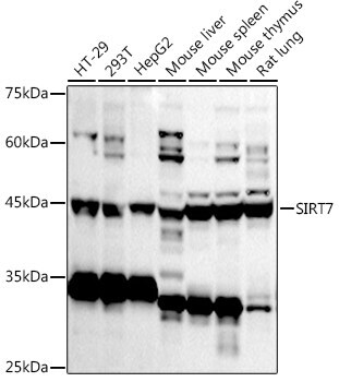
- Western blot analysis of SIRT7 in extracts of various cell lines. Samples were incubated with SIRT7 Polyclonal antibody (Product # PA5-87543) using a dilution of 1:1,000, followed by HRP Goat Anti-Rabbit IgG (H+L) at a dilution of 1:10,000. Lysates/proteins: 25 µg per lane. Blocking buffer: 3% nonfat dry milk in TBST. Detection: ECL Basic Kit. Exposure time: 180s.
Supportive validation
- Submitted by
- Invitrogen Antibodies (provider)
- Main image
- Experimental details
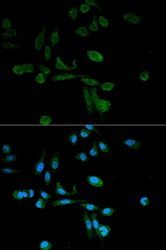
- Immunofluorescence analysis of HeLa cells using SIRT7 Polyclonal antibody (Product # PA5-87543). Blue: DAPI for nuclear staining.

Supportive validation
- Submitted by
- Invitrogen Antibodies (provider)
- Main image
- Experimental details
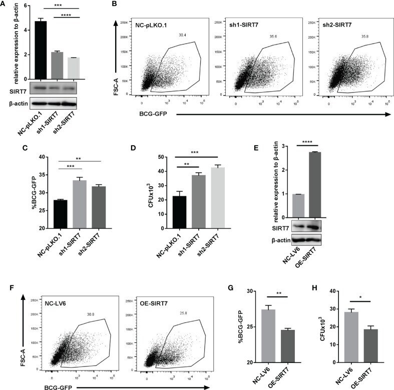
- Figure 3 SIRT7 knockdown increases the risk of Mycobacteria infection, while overexpression of SIRT7 protects cells from Mycobacteria. (A-D) Raw264.7 cells stably expressing scrambled control (NC-pLKO.1) and two independent SIRT7 shRNAs (sh1-SIRT7 and sh2-SIRT7), respectively, were established. SIRT7 expression levels in these cells were measured by quantitative RT-PCR and Western blot analysis (A) . Representative flow cytometry images (B) and percentage (C) of GFP-positive cells were recorded in the scrambled control and SIRT7-knockdown cells 24 h after infection with BCG-GFP (MOI 10:1). (D) Colony-forming unit (CFU) counts in scrambled control and SIRT7-knockdown cells after 48h infection with H37Rv. (E-H) Raw264.7 cells stably overexpressing SIRT7 (OE-SIRT7) and vector control (NC-LV6) were established. SIRT7 expression levels in these cells were measured by quantitative RT-PCR and Western blot analysis (E) . Representative flow cytometry images (F) and percentage (G) of GFP-positive cells were recorded in control and SIRT7-overexpressing cells 24 h after infection with BCG-GFP (MOI 10:1). (H) CFU counts in vector control and SIRT7-overexpressing cells after 48h infected with H37Rv. Data are representative of three independent experiments with similar results and are presented as means +- SD. One way ANOVA was performed in (A, C, D) , Unpaired Student's t-test was used in (E, G, H) . *p < 0.05; **p < 0.01; ***p < 0.001; ****p< 0.0001.