Explore
 Validate
 Learn
Learn Western blot
Antibody data
- Antibody Data
- Antigen structure
- References [0]
- Comments [0]
- Validations
- Western blot [1]
- Immunohistochemistry [1]
- Flow cytometry [1]
Submit
Validation data
Reference
Comment
Report error
- Product number
- MAB5948 - Provider product page

- Provider
- R&D Systems
- Product name
- Human LRP-4 Antibody
- Antibody type
- Monoclonal
- Description
- Protein A or G purified from hybridoma culture supernatant. Detects human LRP-4 in direct ELISAs and Western blots. In direct ELISAs and Western blots, no cross-reactivity with recombinant human (rh) LRP-5 or rhLRP-6 is observed.
- Reactivity
- Human
- Host
- Mouse
- Conjugate
- Unconjugated
- Antigen sequence
O75096- Isotype
- IgG
- Antibody clone number
- 741704
- Vial size
- 100 ug
- Concentration
- LYOPH
- Storage
- Use a manual defrost freezer and avoid repeated freeze-thaw cycles. 12 months from date of receipt, -20 to -70 °C as supplied. 1 month, 2 to 8 °C under sterile conditions after reconstitution. 6 months, -20 to -70 °C under sterile conditions after reconstitution.
No comments: Submit comment
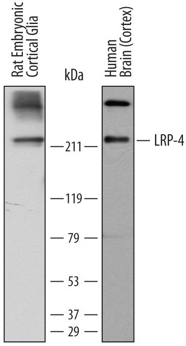
Supportive validation
- Submitted by
- R&D Systems (provider)
- Main image
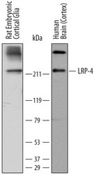
- Experimental details
- Detection of Human and Rat LRP-4 by Western Blot. Western blot shows lysates of rat embryonic cortical glial cells and human brain (cortex) tissue. PVDF membrane was probed with 2 µg/mL of Mouse Anti-Human LRP-4 Monoclonal Antibody (Catalog # MAB5948) followed by HRP-conjugated Anti-Mouse IgG Secondary Antibody (Catalog # HAF007). A specific band was detected for LRP-4 at approximately 220 kDa (as indicated). This experiment was conducted under reducing conditions and using Immunoblot Buffer Group 1.
Supportive validation
- Submitted by
- R&D Systems (provider)
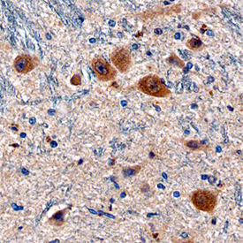
- Main image
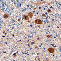
- Experimental details
- LRP-4 in Human Brain. LRP-4 was detected in immersion fixed paraffin-embedded sections of human brain (medulla) using Mouse Anti-Human LRP-4 Monoclonal Antibody (Catalog # MAB5948) at 15 µg/mL overnight at 4 °C. Before incubation with the primary antibody, tissue was subjected to heat-induced epitope retrieval using Antigen Retrieval Reagent-Basic (Catalog # CTS013). Tissue was stained using the Anti-Mouse HRP-DAB Cell & Tissue Staining Kit (brown; Catalog # CTS002) and counter-stained with hematoxylin (blue). Specific staining was localized to cytoplasm of neurons. View our protocol for Chromogenic IHC Staining of Paraffin-embedded Tissue Sections.
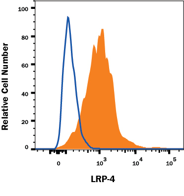
Supportive validation
- Submitted by
- R&D Systems (provider)
- Main image

- Experimental details
- Detection of LRP-4 in U-2 OS Human Cell Line by Flow Cytometry. U-2 OS human osteosarcoma cell line was stained with Mouse Anti-Human LRP-4 Monoclonal Antibody (Catalog # MAB5948, filled histo-gram) or isotype control antibody (Catalog # MAB002, open histogram), followed by Phycoerythrin-conjugated Anti-Mouse IgG Secondary Antibody (Catalog # F0102B). View our protocol for Staining Membrane-associated Proteins.