 Explore
 Validate
 Learn
Learn Immunocytochemistry
Antibody data
- Antibody Data
- Antigen structure
- References [1]
- Comments [0]
- Validations
- Immunocytochemistry [2]
- Flow cytometry [1]
- Other assay [1]
Submit
Validation data
Reference
Comment
Report error
- Product number
- MAB8240 - Provider product page

- Provider
- R&D Systems
- Product name
- Mouse Lgr5/GPR49 Antibody
- Antibody type
- Monoclonal
- Description
- Protein A or G purified from hybridoma culture supernatant. Detects mouse Lgr5/GPR49 in ELISA. Stains mouse Lgr5 transfected cells but not irrelevant transfectants in Flow Cytometry and Immunocytochemistry. This antibody, also known as "RD20", has been found to detect an epitope in C-terminal LRR cap of LGR5 (Ref. 1).
- Reactivity
- Mouse
- Host
- Rat
- Conjugate
- Unconjugated
- Antigen sequence
Q9Z1P4- Isotype
- IgG
- Antibody clone number
- 803420
- Vial size
- 100 ug
- Concentration
- LYOPH
- Storage
- Use a manual defrost freezer and avoid repeated freeze-thaw cycles. 12 months from date of receipt, -20 to -70 °C as supplied. 1 month, 2 to 8 °C under sterile conditions after reconstitution. 6 months, -20 to -70 °C under sterile conditions after reconstitution.
Submitted references Multicolor quantitative confocal imaging cytometry.
Coutu DL, Kokkaliaris KD, Kunz L, Schroeder T
Nature methods 2018 Jan;15(1):39-46
Nature methods 2018 Jan;15(1):39-46
No comments: Submit comment
Supportive validation
- Submitted by
- R&D Systems (provider)
- Main image
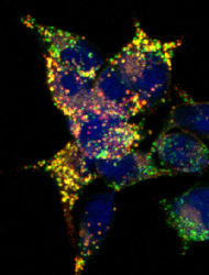
- Experimental details
- Lgr5/GPR49 in HEK293 Human Cell Line Transfected with Mouse Lgf5/GPR49. Lgr5/GPR49 was detected in immersion fixed HEK293 human embryonic kidney cell line transfected with GFP (green) tagged mouse LGR5 using Rat Anti-Mouse Lgr5/GPR49 Monoclonal Antibody (Catalog # MAB8240) at 10 µg/mL for 3 hours at room temperature. Cells were stained using the NorthernLights™ 557-conjugated Anti-Rat IgG Secondary Antibody (red; Catalog # NL013) and counterstained with DAPI (blue). Specific staining was localized to cell surfaces and cytoplasm. View our protocol for Fluorescent ICC Staining of Cells on Coverslips.

- Submitted by
- R&D Systems (provider)
- Main image
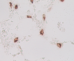
- Experimental details
- Lgr5/GPR49 in Mouse Cells Transfected with Mouse Lgr5/GPR49. Lgr5/GPR49 was detected in immersion fixed MYC tagged mouse transfectants using Rat Anti-Mouse Lgr5/GPR49 Monoclonal Antibody (Catalog # MAB8240). Cells were stained using an anti-Mouse HRP-conjugated secondary antibody (brown). Image courtesy of Dr. Hans Clevers, Hubrecht Institute, The Netherlands.

Supportive validation
- Submitted by
- R&D Systems (provider)
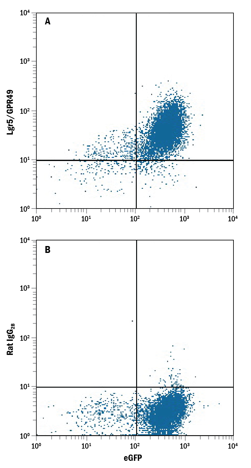
- Main image
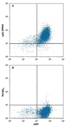
- Experimental details
- Detection of Lgr5/GPR49 in HEK293 Human Cell Line Transfected with Mouse Lgr5/GPR49 and eGFP by Flow Cytometry. HEK293 human embryonic kidney cell line transfected with mouse Lgr5/GPR49 and eGFP was stained with and either (A) Rat Anti-Mouse Lgr5/GPR49 Monoclonal Antibody (Catalog # MAB8240) or (B) Rat IgG2B Isotype Control (Catalog # MAB0061) followed by Allophycocyanin-conjugated Anti-Rat IgG Secondary Antibody (Catalog # F0113).
Supportive validation
- Submitted by
- R&D Systems (provider)
- Main image
- Experimental details
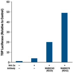
- Mouse Lgr5/GPR49 Antibody Induces Activity. Rat Anti-Mouse Lgr5/GPR49 Monoclonal Antibody (Catalog # MAB8240) induces TOPflash activity in the HEK293 human embryonic kidney cell line stably expressing LGR5 in the presence of Wnt-3a, but in the absence of R-Spondins. (Data courtesy of Dr. Wim de Lau and Dr. Hans Clevers, Hubrecht Institute, The Netherlands. See Reference 1.)
