Explore
 Validate
 Learn
LearnMA5-25644
antibody from Invitrogen Antibodies
Targeting: LGR5
FEX, GPR49, GPR67, HG38
 Western blot
 Immunocytochemistry Immunoprecipitation Immunohistochemistry
 Flow cytometry Other assay
Antibody data
- Antibody Data
- Antigen structure
- References [5]
- Comments [0]
- Validations
- Immunocytochemistry [9]
- Immunoprecipitation [1]
- Immunohistochemistry [9]
- Other assay [7]
Submit
Validation data
Reference
Comment
Report error
- Product number
- MA5-25644 - Provider product page

- Provider
- Invitrogen Antibodies
- Product name
- LGR5 Monoclonal Antibody (OTI2A2)
- Antibody type
- Monoclonal
- Antigen
- Recombinant protein fragment
- Reactivity
- Human, Mouse
- Host
- Mouse
- Isotype
- IgG
- Antibody clone number
- OTI2A2
- Vial size
- 100 μL
- Concentration
- 1 mg/mL
- Storage
- -20°C, Avoid Freeze/Thaw Cycles
Submitted references Characterization of human nasal organoids from chronic rhinosinusitis patients.
Bone Morphogenetic Protein 4 Alleviates DSS-Induced Ulcerative Colitis Through Activating Intestinal Stem Cell by Target ID3.
Clinical Significance of Stem Cell Biomarkers EpCAM, LGR5 and LGR4 mRNA Levels in Lymph Nodes of Colon Cancer Patients.
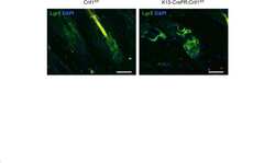
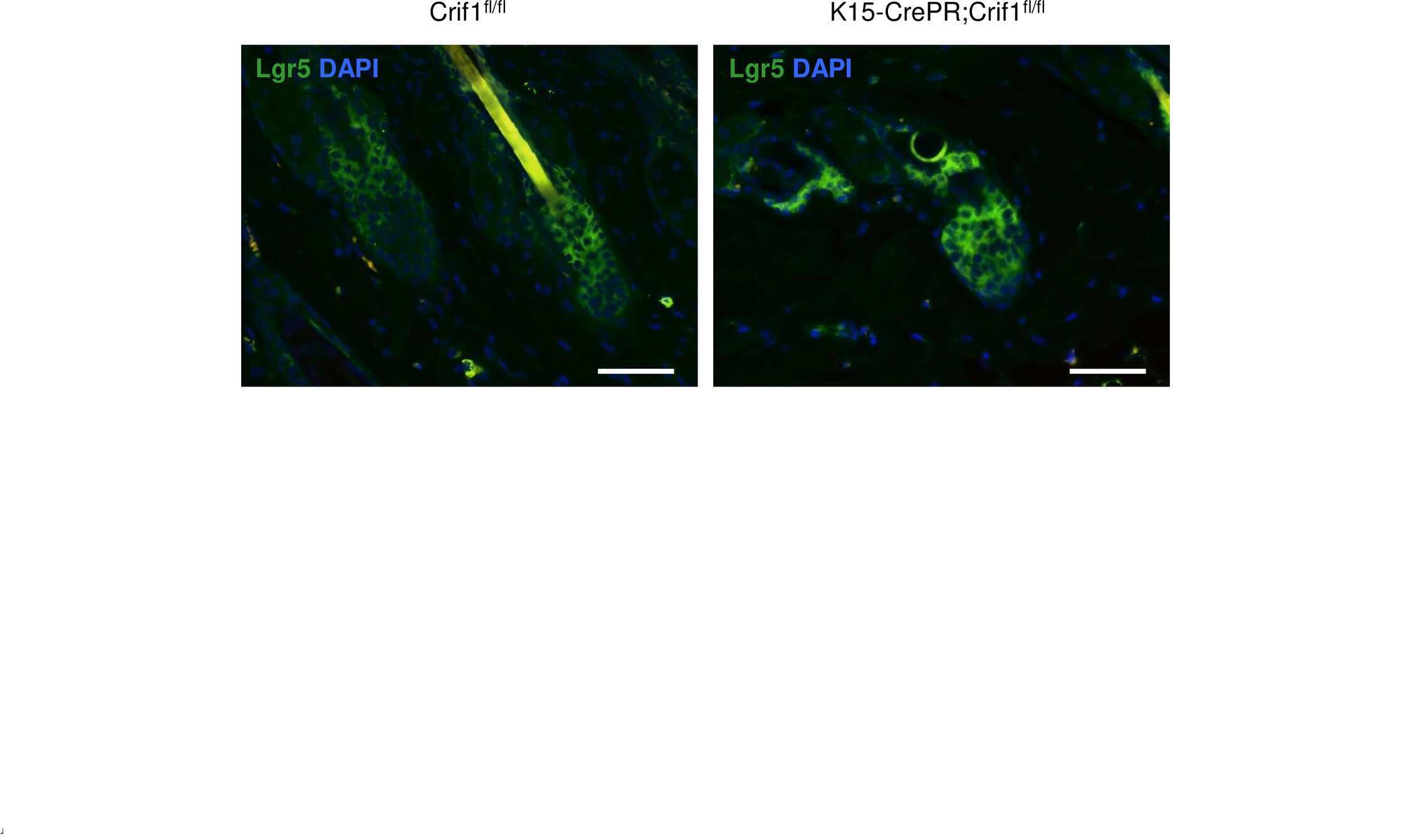
Deficiency of Crif1 in hair follicle stem cells retards hair growth cycle in adult mice.
MUC1-C drives stemness in progression of colitis to colorectal cancer.
Ramezanpour M, Bolt H, Hon K, Shaghayegh G, Rastin H, Fenix KA, Psaltis Alkis J, Wormald PJ, Vreugde S
Biology open 2022 Aug 15;11(8)
Biology open 2022 Aug 15;11(8)
Bone Morphogenetic Protein 4 Alleviates DSS-Induced Ulcerative Colitis Through Activating Intestinal Stem Cell by Target ID3.
Hu L, Xu J, Wang X, Feng L, Zhang C, Wang J, Wang S
Frontiers in cell and developmental biology 2021;9:700864
Frontiers in cell and developmental biology 2021;9:700864
Clinical Significance of Stem Cell Biomarkers EpCAM, LGR5 and LGR4 mRNA Levels in Lymph Nodes of Colon Cancer Patients.
AbdelMageed M, Ismail HTH, Olsson L, Lindmark G, Hammarström ML, Hammarström S, Sitohy B
International journal of molecular sciences 2021 Dec 30;23(1)
International journal of molecular sciences 2021 Dec 30;23(1)
Deficiency of Crif1 in hair follicle stem cells retards hair growth cycle in adult mice.
Shin JM, Ko JW, Choi CW, Lee Y, Seo YJ, Lee JH, Kim CD
PloS one 2020;15(4):e0232206
PloS one 2020;15(4):e0232206
MUC1-C drives stemness in progression of colitis to colorectal cancer.
Li W, Zhang N, Jin C, Long MD, Rajabi H, Yasumizu Y, Fushimi A, Yamashita N, Hagiwara M, Zheng R, Wang J, Kui L, Singh H, Kharbanda S, Hu Q, Liu S, Kufe D
JCI insight 2020 Jun 18;5(12)
JCI insight 2020 Jun 18;5(12)
No comments: Submit comment
Supportive validation
- Submitted by
- Invitrogen Antibodies (provider)
- Main image
- Experimental details
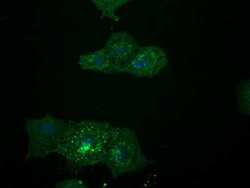
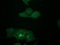
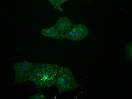
- Immunofluorescent analysis of LGR5 in COS7 cells transfected with a plasmid overexpressing LGR5. Cells were probed with a LGR5 monoclonal antibody (Product # MA5-25644).
- Submitted by
- Invitrogen Antibodies (provider)
- Main image
- Experimental details
- Immunofluorescent analysis of LGR5 in human corneal epithelial cells. Cells were probed with a LGR5 monoclonal antibody (Product # MA5-25644).

- Submitted by
- Invitrogen Antibodies (provider)
- Main image

- Experimental details
- Immunofluorescent analysis of LGR5 in COS7 cells transfected with a plasmid overexpressing LGR5. Cells were probed with a LGR5 monoclonal antibody (Product # MA5-25644).

- Submitted by
- Invitrogen Antibodies (provider)
- Main image

- Experimental details
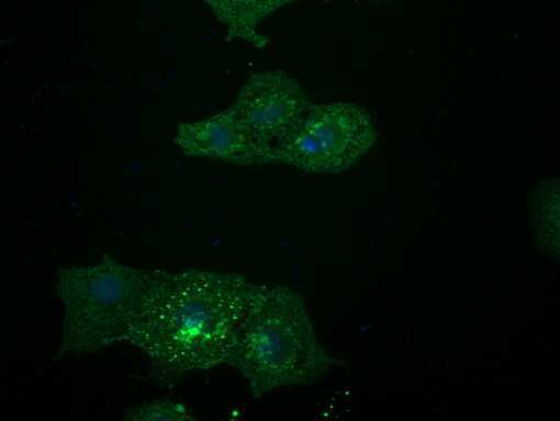
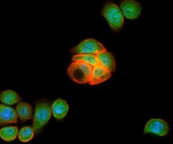
- Immunofluorescent analysis of LGR5 in HT29 cells. Cells were probed with a LGR5 monoclonal antibody (Product # MA5-25644) in green, and actin filaments were probed with TRITC-phalloidin (red), and DAPI (blue) to show the nucleus.

- Submitted by
- Invitrogen Antibodies (provider)
- Main image

- Experimental details
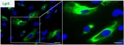
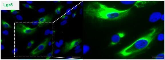
- Immunofluorescent analysis of LGR5 in epihelium stem cells, specifically isolated dental epithelium. Cells were probed with a LGR5 monoclonal antibody (Product # MA5-25644).
- Submitted by
- Invitrogen Antibodies (provider)
- Main image

- Experimental details
- Immunofluorescent analysis of LGR5 in human corneal epithelial cells. Cells were probed with a LGR5 monoclonal antibody (Product # MA5-25644).
- Submitted by
- Invitrogen Antibodies (provider)
- Main image
- Experimental details
- Immunofluorescent analysis of LGR5 in COS7 cells transfected with a plasmid overexpressing LGR5. Cells were probed with a LGR5 monoclonal antibody (Product # MA5-25644).
- Submitted by
- Invitrogen Antibodies (provider)
- Main image
- Experimental details
- Immunofluorescent analysis of LGR5 in HT29 cells. Cells were probed with a LGR5 monoclonal antibody (Product # MA5-25644) in green, and actin filaments were probed with TRITC-phalloidin (red), and DAPI (blue) to show the nucleus.

- Submitted by
- Invitrogen Antibodies (provider)
- Main image
- Experimental details
- Immunofluorescent analysis of LGR5 in epihelium stem cells, specifically isolated dental epithelium. Cells were probed with a LGR5 monoclonal antibody (Product # MA5-25644).

Supportive validation
- Submitted by
- Invitrogen Antibodies (provider)
- Main image
- Experimental details
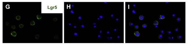
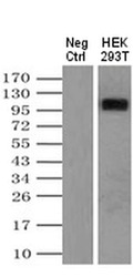
- Immunoprecipitation was performed on HEK293T cells overexpressing LGR5. Antigen-antibody complexes were formed by incubating the sample with 2 µg of LGR5 monoclonal antibody (Product # MA5-25644) antibody and 0.1 mg of goat anti-mouse conjugated magnetic beads overnight. The sample was washed extensively and probed with rabbit anti-DDK polyclonal antibody.

Supportive validation
- Submitted by
- Invitrogen Antibodies (provider)
- Main image
- Experimental details
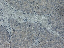
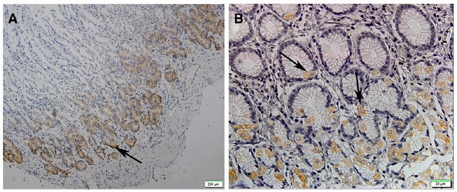
- Immunohistochemistry was performed on paraffin-embedded adenocarcinoma of human breast tissue. To expose target proteins, 10mM citric buffer, pH6.0, 100°C for 10min was used. Following antigen retrieval, tissues were probed with a LGR5 monoclonal antibody (Product # MA5-25644).

- Submitted by
- Invitrogen Antibodies (provider)
- Main image
- Experimental details
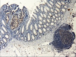
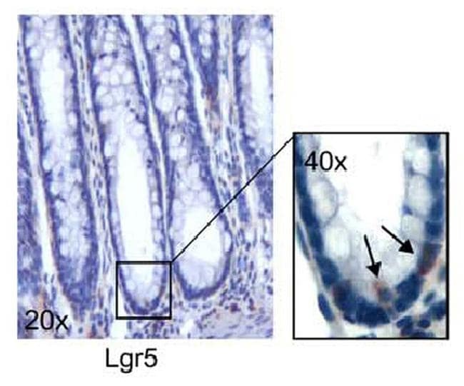
- Immunohistochemistry was performed on paraffin-embedded human colon tissue. To expose target proteins, 10mM citric buffer, pH6.0, 100°C for 10min was used. Following antigen retrieval, tissues were probed with a LGR5 monoclonal antibody (Product # MA5-25644).

- Submitted by
- Invitrogen Antibodies (provider)
- Main image

- Experimental details
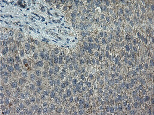
- Immunohistochemistry was performed on paraffin-embedded adenocarcinoma of human colon tissue. To expose target proteins, 10mM citric buffer, pH6.0, 100°C for 10min was used. Following antigen retrieval, tissues were probed with a LGR5 monoclonal antibody (Product # MA5-25644).
- Submitted by
- Invitrogen Antibodies (provider)
- Main image

- Experimental details
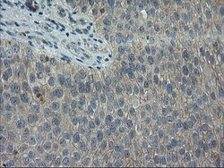
- Immunohistochemistry was performed on paraffin-embedded carcinoma of human thyroid tissue. To expose target proteins, 10mM citric buffer, pH6.0, 100°C for 10min was used. Following antigen retrieval, tissues were probed with a LGR5 monoclonal antibody (Product # MA5-25644).
- Submitted by
- Invitrogen Antibodies (provider)
- Main image
- Experimental details
- Immunohistochemistry was performed on paraffin-embedded carcinoma of human bladder tissue. To expose target proteins, 10mM citric buffer, pH6.0, 100°C for 10min was used. Following antigen retrieval, tissues were probed with a LGR5 monoclonal antibody (Product # MA5-25644).
- Submitted by
- Invitrogen Antibodies (provider)
- Main image
- Experimental details
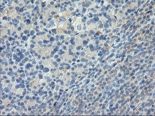
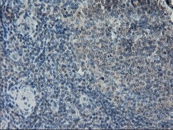
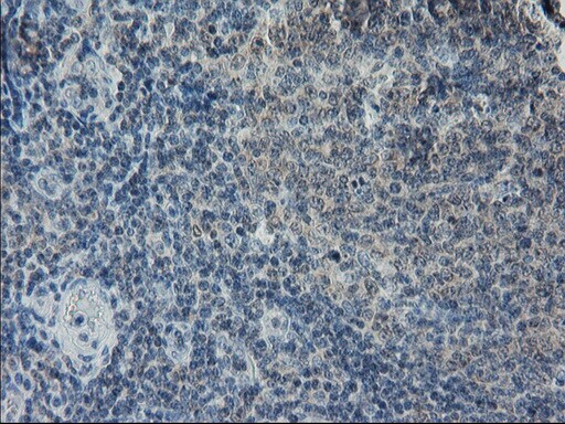
- Immunohistochemistry was performed on paraffin-embedded human lymph node tissue. To expose target proteins, 10mM citric buffer, pH6.0, 100°C for 10min was used. Following antigen retrieval, tissues were probed with a LGR5 monoclonal antibody (Product # MA5-25644).
- Submitted by
- Invitrogen Antibodies (provider)
- Main image
- Experimental details
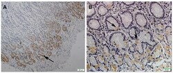
- Immunohistochemical analysis of LGR5 in paraffin-embedded gastric tissue using a LGR5 monoclonal antibody (Product # MA5-25644) at a dilution of 1:100.
- Submitted by
- Invitrogen Antibodies (provider)
- Main image
- Experimental details
- Immunohistochemical analysis of LGR5 in paraffin-embedded gastric mucosa tissue using a LGR5 monoclonal antibody (Product # MA5-25644) at a dilution of 1:100.
- Submitted by
- Invitrogen Antibodies (provider)
- Main image
- Experimental details
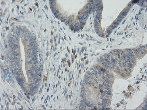
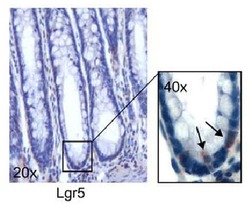
- Immunohistochemical analysis of LGR5 in paraffin-embedded human colon tissue using a LGR5 monoclonal antibody (Product # MA5-25644) at a dilution of 1:200.
Supportive validation
- Submitted by
- Invitrogen Antibodies (provider)
- Main image
- Experimental details
- NULL
- Submitted by
- Invitrogen Antibodies (provider)
- Main image

- Experimental details
- Immunoprecipitation was performed on HEK293T cells overexpressing LGR5. Antigen-antibody complexes were formed by incubating the sample with 2 µg of LGR5 monoclonal antibody (Product # MA5-25644) antibody and 0.1 mg of goat anti-mouse conjugated magnetic beads overnight. The sample was washed extensively and probed with rabbit anti-DDK polyclonal antibody.

- Submitted by
- Invitrogen Antibodies (provider)
- Main image
- Experimental details
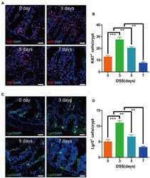
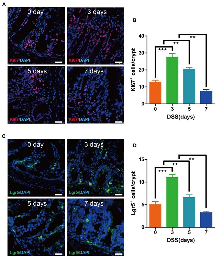
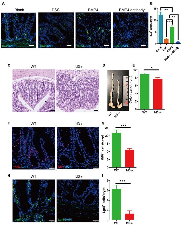
- FIGURE 2 Epithelium cell proliferation and Lgr5 + stem cell maintenance were preserved during the early 3 days of DSS colitis. (A) Immunofluorescence staining for Ki67 in colon tissue from mice treated with DSS at 0, 3, 5, and 7 days (red: Ki67, blue: DAPI). (B) Quantification of Ki67-positive cells per crypt ( n = 5). (C) Immunofluorescence staining for Lgr5 in colon tissue from mice treated with DSS at 0, 3, 5, and 7 days (green: Lgr5, blue: DAPI). (D) Quantification of Lgr5-positive cells per crypt ( n = 5). Data represent mean +- SD with three independent experiments. One-way ANOVA and Bonferroni test for multiple testing was applied in all cases. ** p < 0.01, *** p < 0.001. Scale bar = 25 mum.
- Submitted by
- Invitrogen Antibodies (provider)
- Main image
- Experimental details
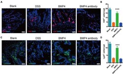
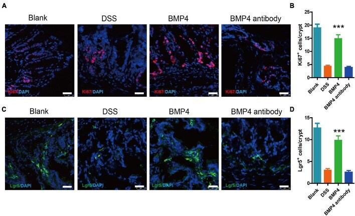
- FIGURE 5 BMP4 maintains epithelium cell proliferation and Lgr5 + ISCs following DSS-induced injury. (A) Immunofluorescence staining for Ki67 in colon tissue from DSS-induced mice treated with saline, BMP4, or anti-BMP4 antibody (red: Ki67, blue: DAPI). (B) Quantification of Ki67-positive cells per crypt ( n = 5). (C) Immunofluorescence staining for Lgr5 in colon tissue from DSS-induced mice treated with saline, BMP4, or anti-BMP4 antibody (green: Lgr5, blue: DAPI). (D) Quantification of Lgr5-positive cells per crypt ( n = 5). Data represent mean +- SD with three independent experiments. One-way ANOVA and Bonferroni tests for multiple testing were applied in all cases. *** p < 0.001. Scale bar = 25 mum.
- Submitted by
- Invitrogen Antibodies (provider)
- Main image
- Experimental details
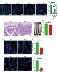
- FIGURE 6 BMP4 ameliorated DSS-induced colitis by target ID3. (A) Immunofluorescence staining for ID3 expression in blank mice, DSS-induced mice treated with saline, BMP4, or anti-BMP4 antibody (green: ID3, blue: DAPI). (B) Quantification of ID3-positive cells per crypt from the colon. (C) Sections of colon tissues stained with H&E from Id3 -/- aged mice and wild-type aged mice. (D,E) Colon length from Id3 -/- aged mice and wild-type aged mice. (F) Immunofluorescence staining of Ki67 in colon tissue from Id3 -/- aged mice and wild-type aged mice. (G) Quantification of Ki67-positive cells per crypt from Id3 -/- aged mice and wild-type aged mice. (H) Immunofluorescence labeling shows the Lgr5-positive cells in tissue from Id3 -/- aged mice and wild-type aged mice. (I) Quantification of Lgr5-positive cells per crypt from Id3 -/- aged mice and wild-type aged mice. Data represent mean +- SD; one-way ANOVA and Bonferroni tests for multiple testing. Student t -test was used for data analyses from Id3 -/- aged mice and wild-type aged mice. * p < 0.05, ** p < 0.01, *** p < 0.001. Scale bar = 25 mum.
- Submitted by
- Invitrogen Antibodies (provider)
- Main image
- Experimental details
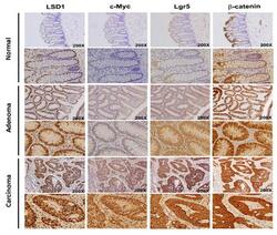
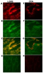
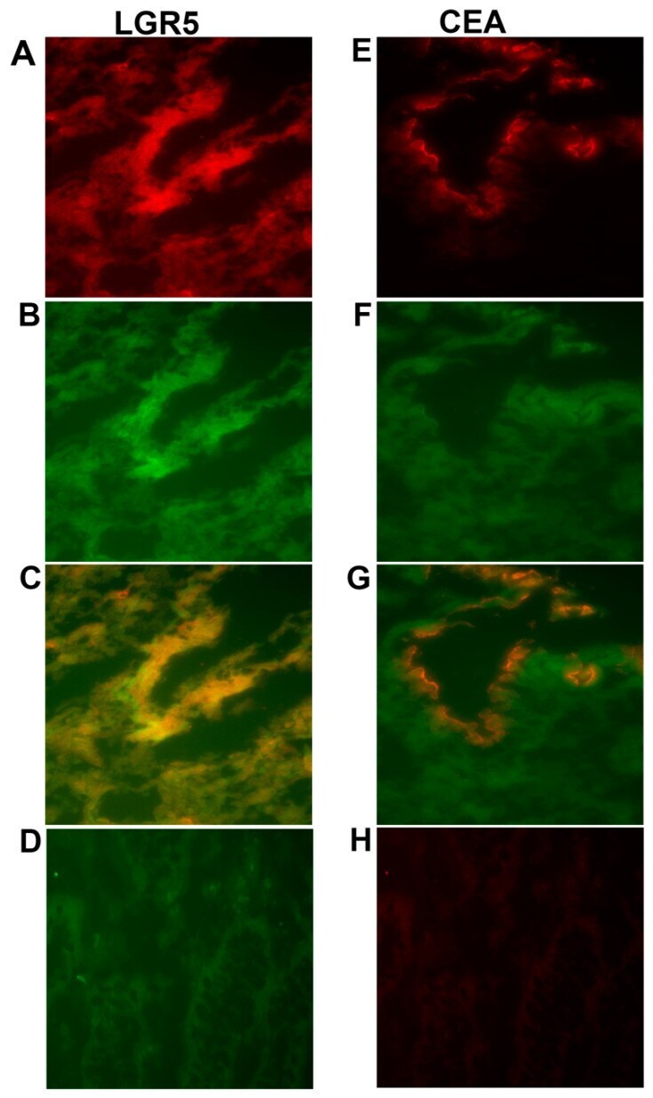
- Figure 3 Two-color immunofluorescence staining of primary colon cancer tissue with anti-LGR5 and BerEP4, and anti-CEA and BerEP4. ( A ) Anti-LGR5, ( E ) Anti-CEA both red color. ( B , F ) BerEP4 mAb, green color. ( C , G ) Overlays giving yellow color of double-stained areas. ( D ) FITC-conjugated mouse IgG; negative control for BerEP4. ( H ) Rabbit IgG; negative control for anti-LGR5 and anti-CEA. Original magnification: x200.
- Submitted by
- Invitrogen Antibodies (provider)
- Main image

- Experimental details
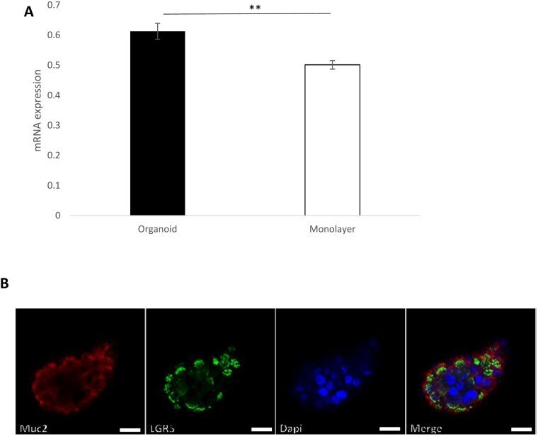
- Fig. 3. LGR5 is highly expressed in nasal organoids. The stem cell marker Lgr5 mRNA expression levels in organoids and monolayer cultures, GAPDH was used as a housekeeping gene and data were normalised to GAPDH. The values are shown as means+-s.e.m., n =5, ** P =0.001 (A). Representative immunofluorescence of 10-12-day-old organoid showing expression of MUC2 (mucus secreting cell, red) and Lgr5 (stem cell marker, green). DAPI stains nuclei blue (B), Scale bar: 20 um and 20x objective.