Explore
 Validate
 Learn
Learn Western blot
 Immunocytochemistry
 Immunoprecipitation
Antibody data
- Antibody Data
- Antigen structure
- References [0]
- Comments [0]
- Validations
- Western blot [1]
- Immunoprecipitation [3]
- Immunohistochemistry [4]
Submit
Validation data
Reference
Comment
Report error
- Product number
- LS-C797459 - Provider product page

- Provider
- LSBio
- Product name
- GPR49 / LGR5 Antibody (aa250-550, clone OTI2A2, Carrier-free) LS-C797459
- Antibody type
- Monoclonal
- Description
- Purified from ascites.
- Reactivity
- Human, Mouse
- Host
- Mouse
- Isotype
- IgG
- Antibody clone number
- OTI2A2
- Storage
- Store at -20°C. Avoid freeze-thaw cycles.
No comments: Submit comment
Enhanced validation
- Submitted by
- LSBio (provider)
- Enhanced method
- Genetic validation
- Main image

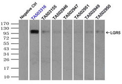
- Experimental details
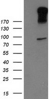
- HEK293T cells were transfected with the pCMV6-ENTRY control. (Left lane) or pCMV6-ENTRY LGR5. (Right lane) cDNA for 48 hrs and lysed
Supportive validation
- Submitted by
- LSBio (provider)
- Enhanced method
- Genetic validation
- Main image
- Experimental details
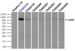
- Immunoprecipitation(IP) of LGR5 by using TrueMab monoclonal anti-LGR5 antibodies. (Negative control: IP without adding anti-LGR5 antibody.). For each experiment, 500ul of DDK tagged LGR5 overexpression lysates. (at 1:5 dilution with HEK293T lysate), 2ug of anti-LGR5 antibody and 20ul. (0.1mg) of goat anti-mouse conjugated magnetic beads were mixed and incubated overnight. After extensive wash to remove any non-specific binding, the immuno-precipitated products were analyzed with rabbit anti-DDK polyclonal antibody.
- Submitted by
- LSBio (provider)
- Main image
- Experimental details
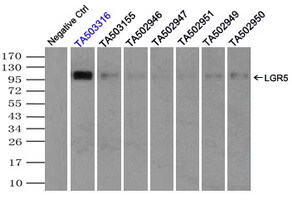
- Immunoprecipitation(IP) of LGR5 by using TrueMab monoclonal anti-LGR5 antibodies. (Negative control: IP without adding anti-LGR5 antibody.). For each experiment, 500ul of DDK tagged LGR5 overexpression lysates. (at 1:5 dilution with HEK293T lysate), 2ug of anti-LGR5 antibody and 20ul. (0.1mg) of goat anti-mouse conjugated magnetic beads were mixed and incubated overnight. After extensive wash to remove any non-specific binding, the immuno-precipitated products were analyzed with rabbit anti-DDK polyclonal antibody.

- Submitted by
- LSBio (provider)
- Main image

- Experimental details
- Immunoprecipitation(IP) of LGR5 by using TrueMab monoclonal anti-LGR5 antibodies. (Negative control: IP without adding anti-LGR5 antibody.). For each experiment, 500ul of DDK tagged LGR5 overexpression lysates. (at 1:5 dilution with HEK293T lysate), 2ug of anti-LGR5 antibody and 20ul. (0.1mg) of goat anti-mouse conjugated magnetic beads were mixed and incubated overnight. After extensive wash to remove any non-specific binding, the immuno-precipitated products were analyzed with rabbit anti-DDK polyclonal antibody.

Supportive validation
- Submitted by
- LSBio (provider)
- Main image
- Experimental details
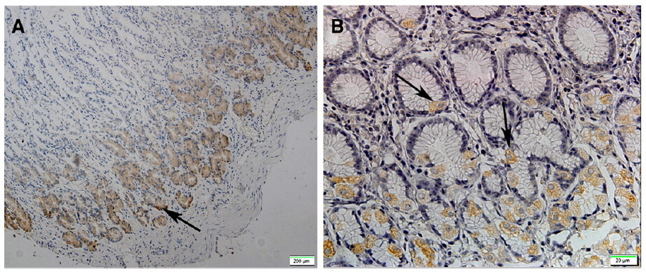
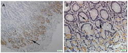
- Figure from citation: Immunohistochemical staining of Lgr5 in normal gastric tissues. The normal gastric tissue, 40×magnified. (A) and 400× magnified. (B). Yellowspots represent the Lgr5 positive cells that are localized only at base of the glands in normal gastric mucosa. The arrows in the pictures show the immunoreactive cells stained in yellow color.

- Submitted by
- LSBio (provider)
- Main image

- Experimental details
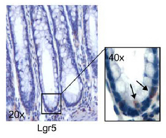
- Figure from citation: Immunohistochemical analysis of Lgr5 expression in human normal colon biopsies; Lgr5+ cells are localized at the crypt base, where bona fide stem-like cells home; magnification is indicated in the boxes. Dilution: 1:200
- Submitted by
- LSBio (provider)
- Main image
- Experimental details
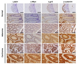
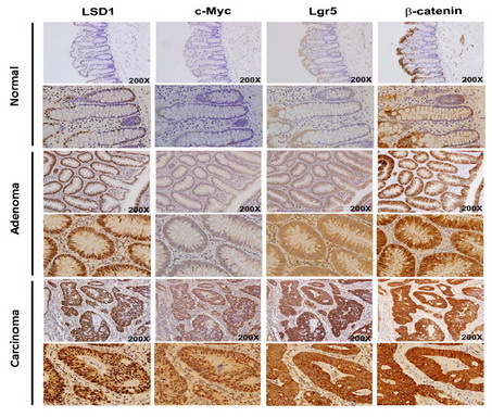
- Figure from citation: Positive correlation between LSD1, c-Myc, ß-catenin and LGR5 expression in human colorectal tumor tissues. Expression levels of LSD1, LGR5, ß-catenin and c-Myc in consecutive sections from normal colon, adenoma and CRC tumor tissues. Dilution: 1:100
- Submitted by
- LSBio (provider)
- Main image

- Experimental details
- Figure from citation: Immunohistochemical staining of Lgr5 in normal gastric tissues. The normal gastric tissue, 40×magnified. (A) and 400× magnified. (B). Yellowspots represent the Lgr5 positive cells that are localized only at base of the glands in normal gastric mucosa. The arrows in the pictures show the immunoreactive cells stained in yellow color.