 Explore
 Validate
 Learn
LearnCF503316
antibody from Invitrogen Antibodies
Targeting: LGR5
FEX, GPR49, GPR67, HG38
 Western blot Immunocytochemistry
 Immunoprecipitation Immunohistochemistry Flow cytometry Other assay
Antibody data
- Antibody Data
- Antigen structure
- References [0]
- Comments [0]
- Validations
- Western blot [1]
- Immunocytochemistry [5]
- Immunohistochemistry [9]
- Flow cytometry [3]
- Other assay [1]
Submit
Validation data
Reference
Comment
Report error
- Product number
- CF503316 - Provider product page

- Provider
- Invitrogen Antibodies
- Product name
- LGR5 Monoclonal Antibody (OTI2A2), TrueMAB™
- Antibody type
- Monoclonal
- Antigen
- Recombinant protein fragment
- Reactivity
- Human, Mouse
- Host
- Mouse
- Isotype
- IgG
- Antibody clone number
- OTI2A2
- Vial size
- 100 µg
- Concentration
- 1 mg/mL
- Storage
- -20° C, Avoid Freeze/Thaw Cycles
No comments: Submit comment
Supportive validation
- Submitted by
- Invitrogen Antibodies (provider)
- Main image

- Experimental details
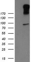
- HEK293T cells were transfected with the pCMV6-ENTRY control (Left lane) or pCMV6-ENTRY LGR5 (RC212825, Right lane) cDNA for 48 hrs and lysed. Equivalent amounts of cell lysates (5 µg per lane) were separated by SDS-PAGE and immunoblotted with anti-LGR5. Positive lysates LY401213 (100 µg) and LC401213 (20 µg) can be purchased separately from OriGene.
Supportive validation
- Submitted by
- Invitrogen Antibodies (provider)
- Main image
- Experimental details
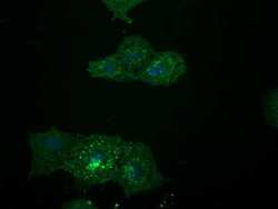
- Anti-LGR5 mouse monoclonal antibody (TA503316) Immunofluorescent staining of COS7 cells transiently transfected by pCMV6-ENTRY LGR5(RC212825).
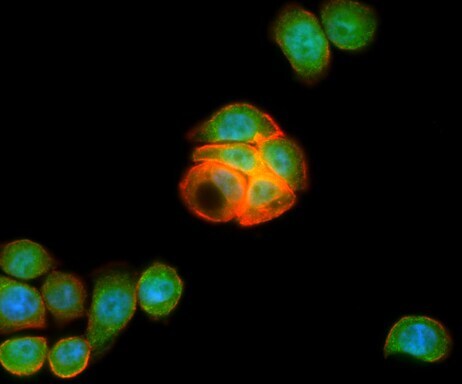
- Submitted by
- Invitrogen Antibodies (provider)
- Main image
- Experimental details
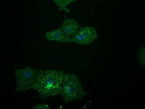
- Anti-LGR5 mouse monoclonal antibody (TA503316) Immunofluorescent staining of COS7 cells transiently transfected by pCMV6-ENTRY LGR5(RC212825).
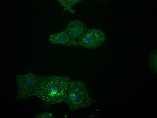
- Submitted by
- Invitrogen Antibodies (provider)
- Main image

- Experimental details
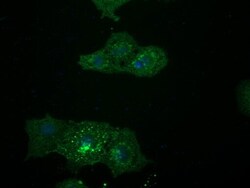
- Immunofluorescent staining of HT29 cells using anti-LGR5 mouse monoclonal antibody (TA503316, green). Actin filaments were labeled with TRITC-phalloidin (red), and nuclear with DAPI (blue).
- Submitted by
- Invitrogen Antibodies (provider)
- Main image
- Experimental details
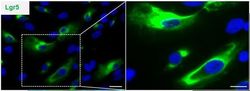
- Figure from citation: Immunocytochemistry of epithelium stem cell phenotype. Isolated dental epithelium following 5-day culture, overwhelmingly expressed Lgr5. Dilution: 1:100 View Citation
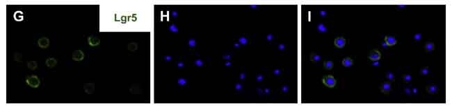
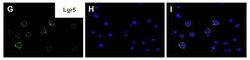
- Submitted by
- Invitrogen Antibodies (provider)
- Main image
- Experimental details
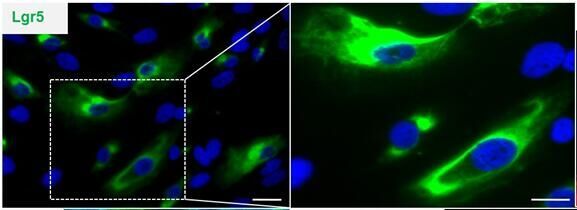
- Figure from citation: [Nature] cells expressing the stem cell marker Lgr5 was detected by immunostaining of hCECs in 2D culture. View CitationPubmed id: 27398792Memo: Human; IF
Supportive validation
- Submitted by
- Invitrogen Antibodies (provider)
- Main image
- Experimental details
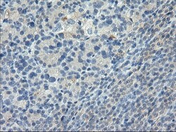
- Immunohistochemical staining of paraffin-embedded Carcinoma of Human thyroid tissue using anti-LGR5 mouse monoclonal antibody. (Heat-induced epitope retrieval by 10mM citric buffer, pH6.0, 100°C for 10min, TA503316)

- Submitted by
- Invitrogen Antibodies (provider)
- Main image
- Experimental details
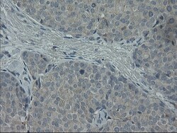
- Immunohistochemical staining of paraffin-embedded Adenocarcinoma of Human breast tissue using anti-LGR5 mouse monoclonal antibody. (Heat-induced epitope retrieval by 10mM citric buffer, pH6.0, 100°C for 10min, TA503316)

- Submitted by
- Invitrogen Antibodies (provider)
- Main image
- Experimental details
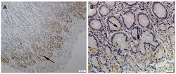
- Figure from citation: Immunohistochemical staining of Lgr5 in normal gastric tissues. The normal gastric tissue, 40×magnified (A) and 400× magnified (B). Yellowspots represent the Lgr5 positive cells that are localized only at base of glands in normal gastric mucosa. The arrows in the pictures show the immunoreactive cells stained in yellow color. View Citation
- Submitted by
- Invitrogen Antibodies (provider)
- Main image
- Experimental details
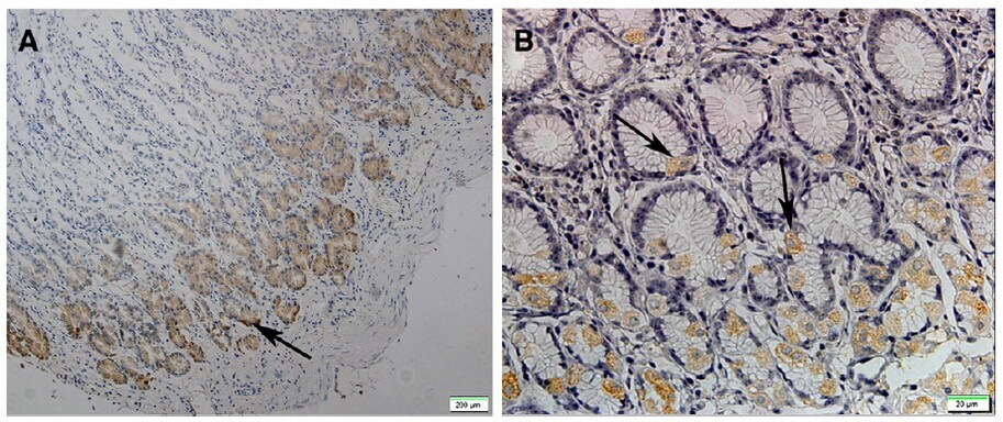
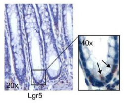
- Figure from citation: Immunohistochemical analysis of Lgr5 expression in human normal colon biopsies; Lgr5+ cells are localized at the crypt base, where bona fide stem-like cells home; magnification is indicated in the boxes. Dilution: 1:200 View Citation

- Submitted by
- Invitrogen Antibodies (provider)
- Main image
- Experimental details
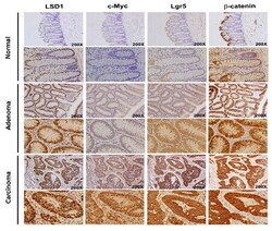
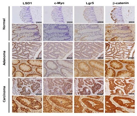
- Figure from citation: Positive correlation between LSD1, c-Myc, beta-catenin and LGR5 expression in human colorectal tumor tissues. Expression levels of LSD1, LGR5, beta-catenin and c-Myc in consecutive sections from normal colon, adenoma and CRC tumor tissues. Dilution: 1:100 View Citation
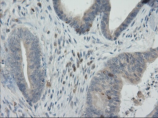
- Submitted by
- Invitrogen Antibodies (provider)
- Main image

- Experimental details
- Immunohistochemical staining of paraffin-embedded Carcinoma of Human bladder tissue using anti-LGR5 mouse monoclonal antibody. (Heat-induced epitope retrieval by 10mM citric buffer, pH6.0, 100°C for 10min, TA503316)
- Submitted by
- Invitrogen Antibodies (provider)
- Main image
- Experimental details
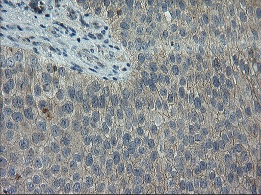
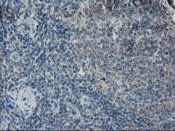
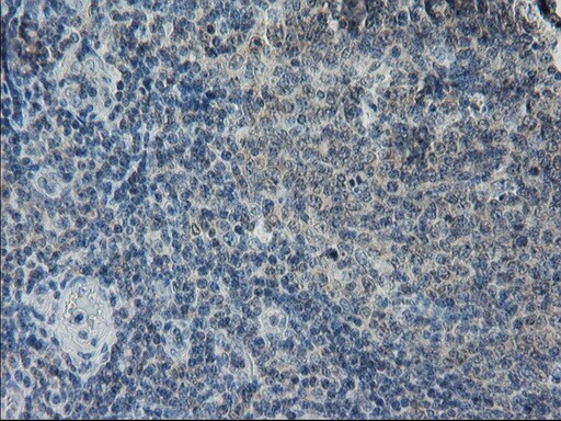
- Immunohistochemical staining of paraffin-embedded human lymph node tissue within the normal limits using anti-LGR5 mouse monoclonal antibody. (Heat-induced epitope retrieval by 10mM citric buffer, pH6.0, 100°C for 10min, TA503316)
- Submitted by
- Invitrogen Antibodies (provider)
- Main image
- Experimental details
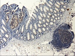
- Immunohistochemical staining of paraffin-embedded human colon tissue within the normal limits using anti-LGR5 mouse monoclonal antibody. (Heat-induced epitope retrieval by 10mM citric buffer, pH6.0, 100°C for 10min, TA503316)

- Submitted by
- Invitrogen Antibodies (provider)
- Main image

- Experimental details
- Immunohistochemical staining of paraffin-embedded Adenocarcinoma of Human colon tissue using anti-LGR5 mouse monoclonal antibody. (Heat-induced epitope retrieval by 10mM citric buffer, pH6.0, 100°C for 10min, TA503316)
Supportive validation
- Submitted by
- Invitrogen Antibodies (provider)
- Main image
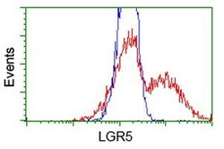
- Experimental details
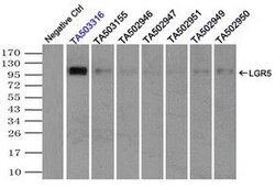
- HEK293T cells transfected with either RC212825 overexpress plasmid (Red) or empty vector control plasmid (Blue) were immunostained by anti-LGR5 antibody (TA503316), and then analyzed by flow cytometry.
- Submitted by
- Invitrogen Antibodies (provider)
- Main image
- Experimental details
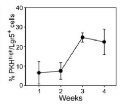
- Figure from citation: Cytofluorometric analysis of Lgr5 expression by PKH high cells over 4 weeks of culture. Data are expressed as percent mean values (6 SD) of consecutive experiments. Dilution: 1:400 View Citation

- Submitted by
- Invitrogen Antibodies (provider)
- Main image
- Experimental details
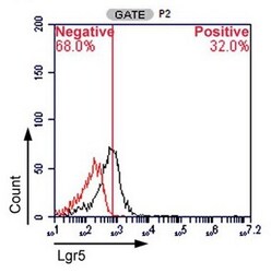
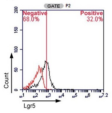
- Figure from citation: Flow cytometry of SB cells. The G4 region was further divided into P1 and P2. Lgr5, a stem cell marker, was expressed by 32% of P2 population. Black: staining with Lgr5; red: staining with the isotype control. View Citation
Supportive validation
- Submitted by
- Invitrogen Antibodies (provider)
- Main image
- Experimental details
- Immunoprecipitation(IP) of LGR5 by using TrueMab monoclonal anti-LGR5 antibodies (Negative control: IP without adding anti-LGR5 antibody.). For each experiment, 500ul of tagged LGR5 overexpression lysates (at 1:5 dilution with HEK293T lysate), 2 µg of anti-LGR5 antibody and 20ul (0.1mg) of goat anti-mouse conj µgated magnetic beads were mixed and incubated overnight. After extensive wash to remove any non-specific binding, the immuno-precipitated products were analyzed with rabbit anti-DDK polyclonal antibody.
