Explore
 Validate
 Learn
Learn Western blot
 Immunocytochemistry
Antibody data
- Antibody Data
- Antigen structure
- References [3]
- Comments [0]
- Validations
- Immunocytochemistry [1]
- Immunohistochemistry [1]
Submit
Validation data
Reference
Comment
Report error
- Product number
- AMAb90679 - Provider product page

- Provider
- Atlas Antibodies
- Proper citation
- Atlas Antibodies Cat#AMAb90679, RRID:AB_2665629
- Product name
- Anti-SATB2
- Antibody type
- Monoclonal
- Description
- Monoclonal Antibody against Human SATB2, Clone ID: CL0320, Gene description: SATB homeobox 2, Alternative Gene Names: FLJ21474, KIAA1034, Validated applications: WB, IHC, ICC, Uniprot ID: Q9UPW6, Storage: Store at +4°C for short term storage. Long time storage is recommended at -20°C.
- Reactivity
- Human, Rat
- Host
- Mouse
- Conjugate
- Unconjugated
- Isotype
- IgG
- Antibody clone number
- CL0320
- Vial size
- 100 µl
- Concentration
- 0.1 mg/ml
- Storage
- Store at +4°C for short term storage. Long time storage is recommended at -20°C.
- Handling
- The antibody solution should be gently mixed before use.
Submitted references Delineating the intra-patient heterogeneity of molecular alterations in treatment-naïve colorectal cancer with peritoneal carcinomatosis.
Prognostic and treatment predictive significance of SATB1 and SATB2 expression in pancreatic and periampullary adenocarcinoma.
SATB1 is an independent prognostic factor in radically resected upper gastrointestinal tract adenocarcinoma.
Siesing C, Petersson A, Ulfarsdottir T, Chattopadhyay S, Nodin B, Eberhard J, Brändstedt J, Syk I, Gisselsson D, Jirström K
Modern pathology : an official journal of the United States and Canadian Academy of Pathology, Inc 2022 Jul;35(7):979-988
Modern pathology : an official journal of the United States and Canadian Academy of Pathology, Inc 2022 Jul;35(7):979-988
Prognostic and treatment predictive significance of SATB1 and SATB2 expression in pancreatic and periampullary adenocarcinoma.
Elebro J, Heby M, Gaber A, Nodin B, Jonsson L, Fristedt R, Uhlén M, Jirström K, Eberhard J
Journal of translational medicine 2014 Oct 17;12:289
Journal of translational medicine 2014 Oct 17;12:289
SATB1 is an independent prognostic factor in radically resected upper gastrointestinal tract adenocarcinoma.
Hedner C, Gaber A, Korkocic D, Nodin B, Uhlén M, Kuteeva E, Johannesson H, Jirström K, Eberhard J
Virchows Archiv : an international journal of pathology 2014 Dec;465(6):649-59
Virchows Archiv : an international journal of pathology 2014 Dec;465(6):649-59
No comments: Submit comment
Supportive validation
- Submitted by
- Atlas Antibodies (provider)
- Main image

- Experimental details
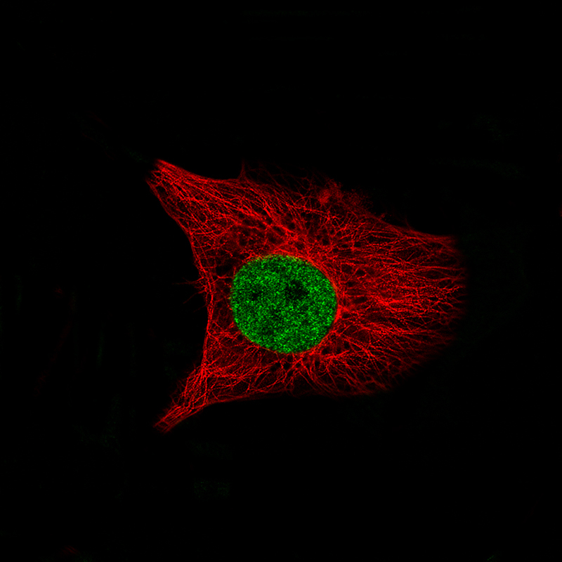
- Immunofluorescence staining of U-138 MG cells using the Anti-SATB2 monoclonal antibody, showing specific staining in the nucleoplasm in green. Microtubule- and nuclear probes are visualized in red and blue, respectively (where available).
- Sample type
- Human
Supportive validation
- Submitted by
- Atlas Antibodies (provider)
- Enhanced method
- Orthogonal validation
- Main image
- Experimental details
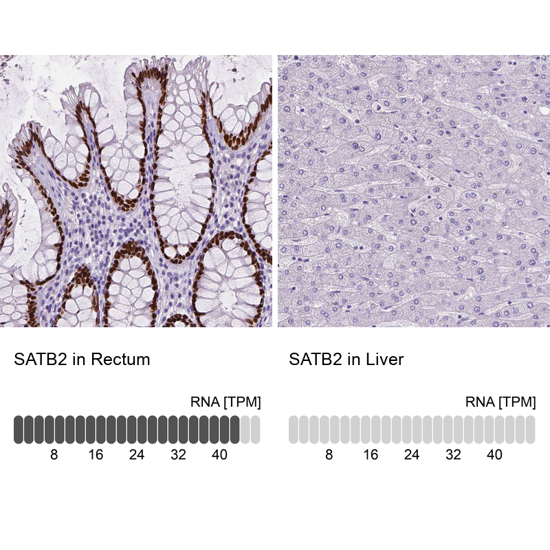
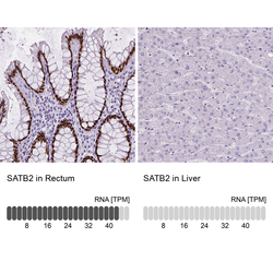
- Immunohistochemistry analysis in human rectum and liver tissues using AMAb90679 antibody. Corresponding SATB2 RNA-seq data are presented for the same tissues.
- Sample type
- Human
- Protocol
- Protocol